Japanese
English
- 有料閲覧
- Abstract 文献概要
- 1ページ目 Look Inside
I.はじめに
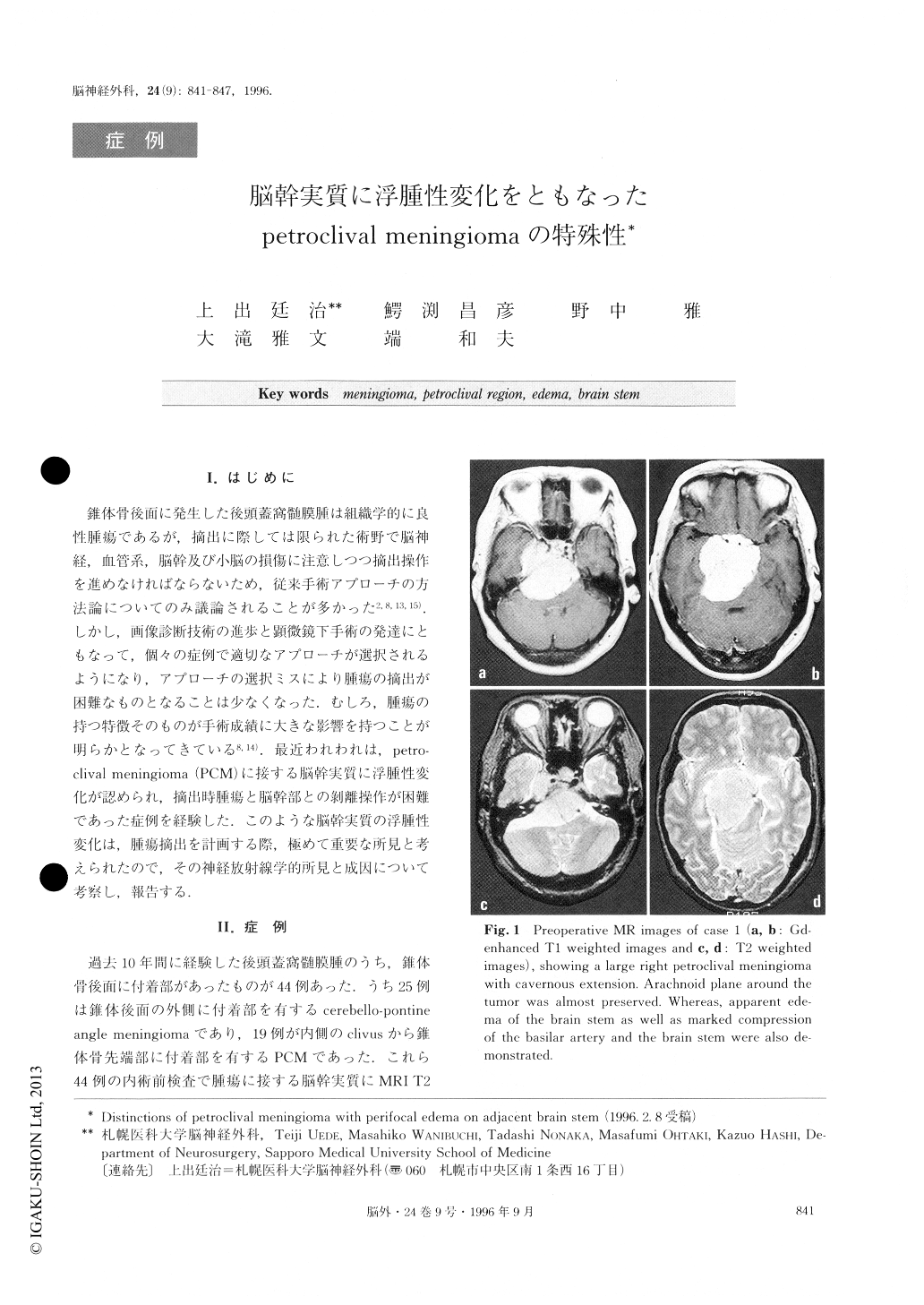
錐体骨後面に発生した後頭蓋窩髄膜腫は組織学的に良性腫瘍であるが,摘出に際しては限られた術野で脳神経,血管系,脳幹及び小脳の損傷に注意しつつ摘出操作を進めなければならないため,従来手術アプローチの方法論についてのみ議論されることが多かった2,8,13,15).しかし,画像診断技術の進歩と顕微鏡下手術の発達にともなって,個々の症例で適切なアプローチが選択されるようになり,アプローチの選択ミスにより腫瘍の摘出が困難なものとなることは少なくなった.むしろ,腫瘍の持つ特徴そのものが手術成績に大きな影響を持つことが明らかとなってきている8,14).最近われわれは,petro—clival meningioma(PCM)に接する脳幹実質に浮腫性変化が認められ,摘出時腫瘍と脳幹部との剥離操作が困難であった症例を経験した.このような脳幹実質の浮腫性変化は,腫瘍摘出を計画する際,極めて重要な所見と考えられたので,その神経放射線学的所見と成因について考察し,報告する.
Although the most frequent benign tumor of the cen-tral nervous system, meningioma may be associated with extensive peritumoral edema. Whereas, peritumoral edema in the brain stem along the tumor in the infra-tentorial region has not been given sufficient recogni-tion. Among 44 meningiomas attached to the petrous bone, 25 cerebellopontine angle meningiomas and 17 petroclival meningiomas, peritumoral edema in the brain stem along the tumor were clearly demonstrated on T2 weighted images of MRI in three petroclival meningiomas (6.8% of all clival and 17.6% of petrocli-val meningiomas). Attempts were made to surgically remove all of these tumors. However, during surgeryan arachnoid/pial layer between the tumor and brain stem was destroyed completely and small perforating arteries were found to be encased in the tumor at the level of the associated edema. So surgical dissection of the tumors from the brain stem in those regions was quite difficult. By attempting radical removal of the tumor in the first case, even meticulous dissections caused direct surgical damage in the brain stem due to the obliteration of small perforating vessels. In the second case, a thin layer of the tumor remnant beside the brain stem was left intentionally, but a severe dam-age of the brain stem also occurred due to the oblitera-tion of small perforating vessels. In the third case, only bulk reduction of the tumor was attempted to minimize the mass effect to the brain stem and the tumor located beside the brain stem with edema and encasing the cranial nerves and the perforating arteries from the vertebrobasilar artery was left untouched. This surgical attempt caused a transient worsening of a swallowing disturbance, but was not associated with the brain stem damage in this case.
In meningiomas attached to the clivus, existence of associated peritumoral edema in the adjacent brain stem on T2 weighted images on MRI may be caused by the destruction of an arachnoid/pial layer and the encasement of small perforating vessels. The attempt at tumor dissection from the brain stem at the level of the edema was very difficult and led to serious surgical complications due to direct damage to the brain stem. In such cases, the surgery should aim at achieving a simple bulk reduction and the tumor beside the brain stem with edema should be left untouched.
Copyright © 1996, Igaku-Shoin Ltd. All rights reserved.