Japanese
English
- 有料閲覧
PPV購入案内
1,320円 (1,200円+税10%) こちらは、779ページから786ページの文献です。購入には医書.jp本会員登録が必要です。
会員登録完了後に改めて上記「購入サイトへ」を選択してください。
または「購入サイトへ」選択後に「購入ログイン」、「新規会員登録」の順に進んでください。
会員登録について詳しくはをご確認ください。
- Abstract 文献概要
- 1ページ目 Look Inside
I.はじめに
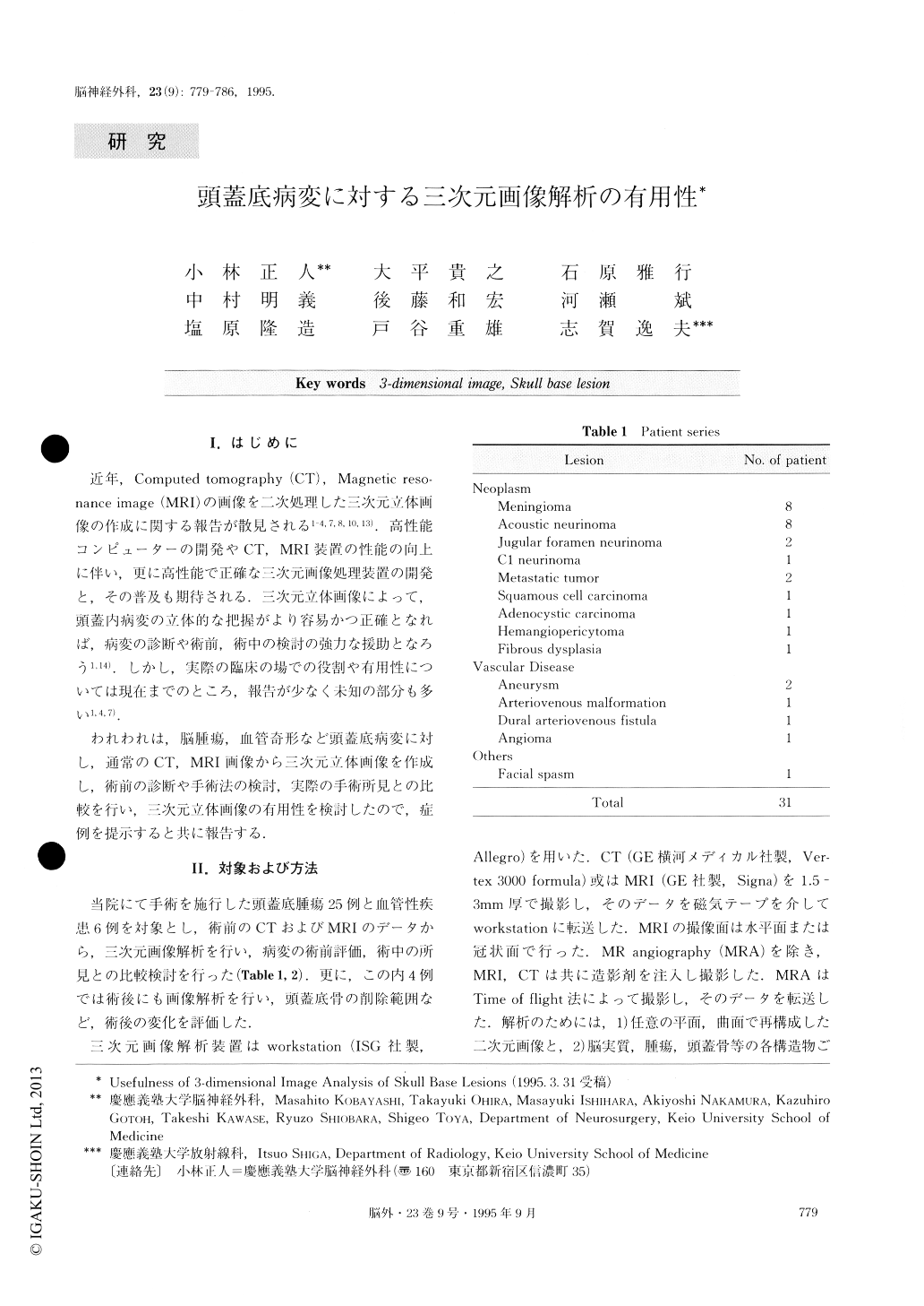
近年,Computed tomography(CT),Magnetic reso—nance image(MRI)の画像を二次処理した三次元立体画像の作成に関する報告が散見される1-4,7,8,10,13).高性能コンピューターの開発やCT,MRI装置の性能の向上に伴い,更に高性能で正確な三次元画像処理装置の開発と,その普及も期待される.三次元立体画像によって,頭蓋内病変の立体的な把握がより容易かつ正確となれば,病変の診断や術前,術中の検討の強力な援助となろう1,14).しかし,実際の臨床の場での役割や有用性については現在までのところ,報告が少なく未知の部分も多い1,4,7).
われわれは,脳腫瘍,血管奇形など頭蓋底病変に対し,通常のCT,MRI画像から三次元立体画像を作成し,術前の診断や手術法の検討,実際の手術所見との比較を行い,三次元立体画像の有用性を検討したので,症例を提示すると共に報告する.
We have evaluated three-dimensional (3D) images of the skull base lesions for planning cranial base surgery. Fifty 3D images were reconstructed from computed tomographies (CT), and/or magnetic resonance (MR) images or MR angiographic images of 30 patients with skull base lesions. These images have provided useful information for pre-operative evaluation. The 3D image reconstructed from CT provides clear information con-cerning the bone. Conversely, the 3D image from MR images demonstrates soft tissue very clearly, and that from MR angiography provides a detailed description of the vasculature. For skull base lesions, it is essential to evaluate 3D images from the different modalities, especially CT scan and MR image.
Copyright © 1995, Igaku-Shoin Ltd. All rights reserved.