Japanese
English
- 有料閲覧
PPV購入案内
1,320円 (1,200円+税10%) こちらは、781ページから784ページの文献です。購入には医書.jp本会員登録が必要です。
会員登録完了後に改めて上記「購入サイトへ」を選択してください。
または「購入サイトへ」選択後に「購入ログイン」、「新規会員登録」の順に進んでください。
会員登録について詳しくはをご確認ください。
- Abstract 文献概要
- 1ページ目 Look Inside
I.はじめに
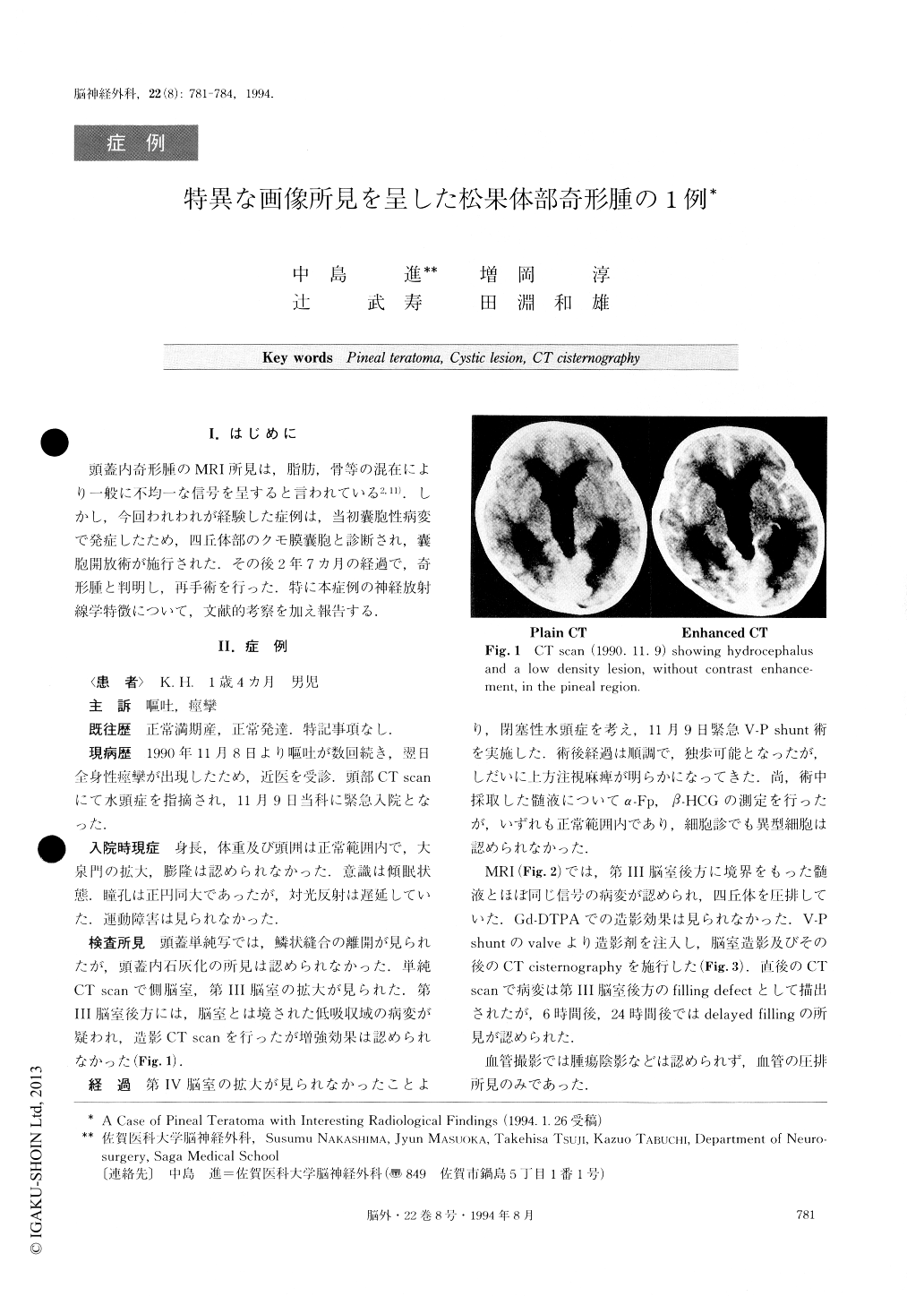
頭蓋内奇形腫のMRI所見は,脂肪,骨等の混在により一般に不均一な信号を呈すると言われている2,11).しかし,今回われわれが経験した症例は,当初嚢胞性病変で発症したため,四丘体部のクモ膜嚢胞と診断され,嚢胞開放術が施行された.その後2年7カ月の経過で,奇形腫と判明し,再手術を行った.特に本症例の神経放射線学特徴について,文献的考察を加え報告する.
A rare case of mature pineal teratoma with interest-ing radiological findings in a 16-month-old infant is re-ported. The patient was referred to our clinic because of generalized convulsions. A CT scan showed marked hydrocephalus and a low density mass lesion without contrast enhancement in the pineal region. A CT cister-nography demonstrated the lesion as a filling defect area on the image produced immediately after the emergent V-P shunt, and a filling area 24 hours later. The lesion was of signal intensity on T-1 weighted MR image and of high signal intensity on T-2 weighted MR image, equal to CSF intensity. These radiological find-ings were compatible with an arachnoid cyst in the quadrigeminal cistern, so we performed the excision of both anterior and posterior cyst walls using the occipit-al tentorial approach. However, the histology of the cyst wall was not compatible with that of an arachnoid cyst, showing neuroepithelial-like cell lining with posi-tive staining for cytokeratine. The postoperative follow-up on MRI was continued for 31 months. An MRI per-formed 9 days after the first operation showed, com-pared with the gray matter, iso-signal intensity area and high signal intensity area on T-1 weighted image. At first, we thought this was because of bleeding of the pineal gland brought on by the operative maneuver. However, the mixed intensity lesion shown on T-1 weighted images gradually expanded and distorted, and finally showed the typical MR images for a teratoma. The operative findings using the occipital transtentorial approach were typical for a teratoma. It contained hair and cartilaginous as well as adipose tissues, and the teratoma was totally removed.
We stress that a mature teratoma should be regarded as a possibility for differential diagnosis when a cystic lesion in the quadrigeminal cistern is encountered.
Copyright © 1994, Igaku-Shoin Ltd. All rights reserved.