Japanese
English
- 有料閲覧
PPV購入案内
1,320円 (1,200円+税10%) こちらは、589ページから592ページの文献です。購入には医書.jp本会員登録が必要です。
会員登録完了後に改めて上記「購入サイトへ」を選択してください。
または「購入サイトへ」選択後に「購入ログイン」、「新規会員登録」の順に進んでください。
会員登録について詳しくはをご確認ください。
- Abstract 文献概要
- 1ページ目 Look Inside
I.はじめに
脊髄空洞症の原因としてキアリ奇形,癒着性クモ膜炎,外傷,脊髄腫瘍などが知られている3).MRI普及後,多発性硬化症にも空洞病変を伴うことが散見され注目されるようになってきた.今回われわれは脊髄空洞病変をともなった多発性硬化症の症例を経験したので文献的考察を加え報告する.
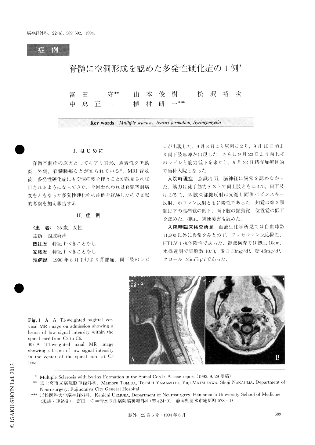
A 35-year-female developed tetraparesis following a period of back pain, urinary retension and paraparesis. She was admitted to our hospital on September 22, 1990. Neurological examination revealed tetraparesis with a sensory level at C3. Deep tendon reflexes were hyperactive in all four extremities with bilateral Hoff-mann and Babinski reflexes. Spinal MR imaging de-monstrated cavity formation within the cervical cord, extending from C2 to C6 without any anomalies at the craniocervical junction including Chiari malformation.The cavity was not enhanced with Gd-DTPA. Cere-brospinal fluid (CSF) examination revealed no abnor-mality. Although a syringo-subarachnoid shunt was performed on October 18, 1990, her symptoms did not improve. On December 17, 1990, she developed optic neuritis in the right eye and her paraparesis deterio-rated. Delayed metrizamide CT scans showed another syrinx from Th3 to Th9. Repeated CSF contained 37 cells/mm3, protein 88 mg% and sugar 49 mg%. CSF oli-goclonal bands were present and the CSF myelin basic protein was 6.6 ng/ml (normal<4). A pulse therapy of steroid improved her vision and paraparesis. However, she developed paraparesis again on October 29, 1991. A brain T2-weighted MR image demonstrated multiple periventricular high signal intensity spots. Her para-paresis improved again with steroid. MR images in September, 1992 revealed a cord atrophy and dis-appearance of the cavity. Based on these clinical courses and radiological findings, a definite diagnosis of multiple sclerosis (MS) was made. We think MS may have caused the syringomyelia.
Copyright © 1994, Igaku-Shoin Ltd. All rights reserved.