Japanese
English
- 有料閲覧
PPV購入案内
1,320円 (1,200円+税10%) こちらは、1189ページから1194ページの文献です。購入には医書.jp本会員登録が必要です。
会員登録完了後に改めて上記「購入サイトへ」を選択してください。
または「購入サイトへ」選択後に「購入ログイン」、「新規会員登録」の順に進んでください。
会員登録について詳しくはをご確認ください。
- Abstract 文献概要
- 1ページ目 Look Inside
I.はじめに
頭蓋内の神経鞘腫の多くは聴神経に発生し,次いで三叉神経にみられるが,前頭蓋底に発生する神経鞘腫,すなわちsubfrontal schwannomaは極めて稀で,現在までに11例1,6,8,14,15,20,22,23,25-27)の報告があるに過ぎない.今回,われわれは鼻腔,篩骨洞から前頭蓋底に至るsubfrontal schwannomaの1例を経験したので報告する.
We encountered a rare case of subfrontal schwannoma. A 55-year-old woman had received resection of a left frontal tumor because of hyposmia, at the age of 28 years. on June 10, 1989, she was admitted with the chief complaint of progressive contraction of visual field. Neu-rologic examination revealed anosmia, impaired vision and concentric contraction of visual field. Fundoscopy showed optic atrophy.
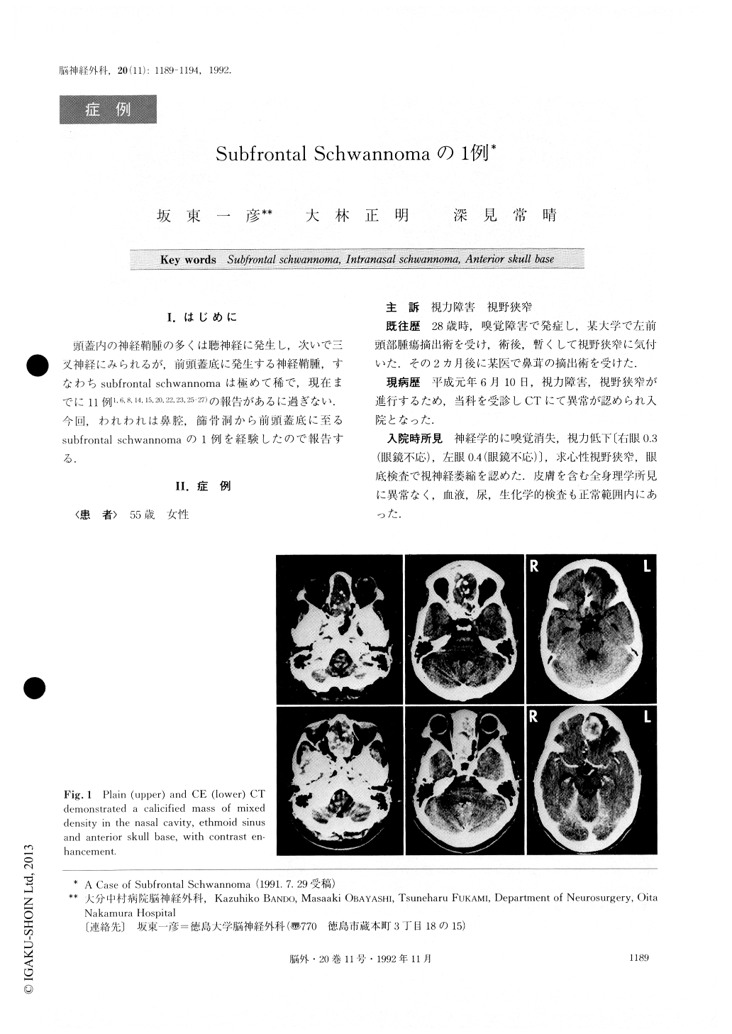
CT examination demonstrated a calcified mass of mixed density which was occupying her nasal cavity, ethmoid sinus and anterior skull base. The lesion was en-hanced with contrast medium. MRI clearly depicted the extension of the lesion and a low signal intensity area in the left frontal lobe as a postoperative scar. Angiography showed hypovascularity.
The tumor was totally removed by bifrontal cra-niotomy on August 22, 1989. Infiltration into the brain or compression of the optic nerve was not detected. The dura on the cranial base side was damaged and lost by infiltration of the tumor, normal olfactory bulb was not able to be identified, and the cribrifrom plate was broken. The anterior skull base was reconstituted bycovering the dttral defect with cadaveric dura and the bony defect with a pericranium.
HE staining showed Antoni A&B types of schwanno-ma. Postoperative course was uneventful. In this case, it is most likely that a remnant of the tumor resected when she was 28 years old had developed subfrontal schwannoma a long time after the operation, although the histological type at that time was unknown. It is also possible that a primary tumor in the nasal cav-ity or paranasal sinus may have extended into the cra-nium. Of 11 reports of subfrontal schwannoma, 2 reports describe extracranial extension to the paranasal sinus.Primary schwannoma in the nasal cavity or paranasal sinus was reported to extend to the anterior skull base in 3 cases, and to the middle cranial fossa in 4 cases of 87 cases reported.
In our case, the possible subfrontal schwannoma is assumed to have originated from 1) the meningeal branch of the trigeminal nerve or the anterior ethmoid nerve, 2) aberration, or 3) the perivascular nerve plexus in the pia mater. However, the definite orgin was not able to be lo-cated owing to the previous operation.
Copyright © 1992, Igaku-Shoin Ltd. All rights reserved.