Japanese
English
- 有料閲覧
PPV購入案内
1,320円 (1,200円+税10%) こちらは、570ページから576ページの文献です。購入には医書.jp本会員登録が必要です。
会員登録完了後に改めて上記「購入サイトへ」を選択してください。
または「購入サイトへ」選択後に「購入ログイン」、「新規会員登録」の順に進んでください。
会員登録について詳しくはをご確認ください。
- Abstract 文献概要
- 1ページ目 Look Inside
- 参考文献 Reference
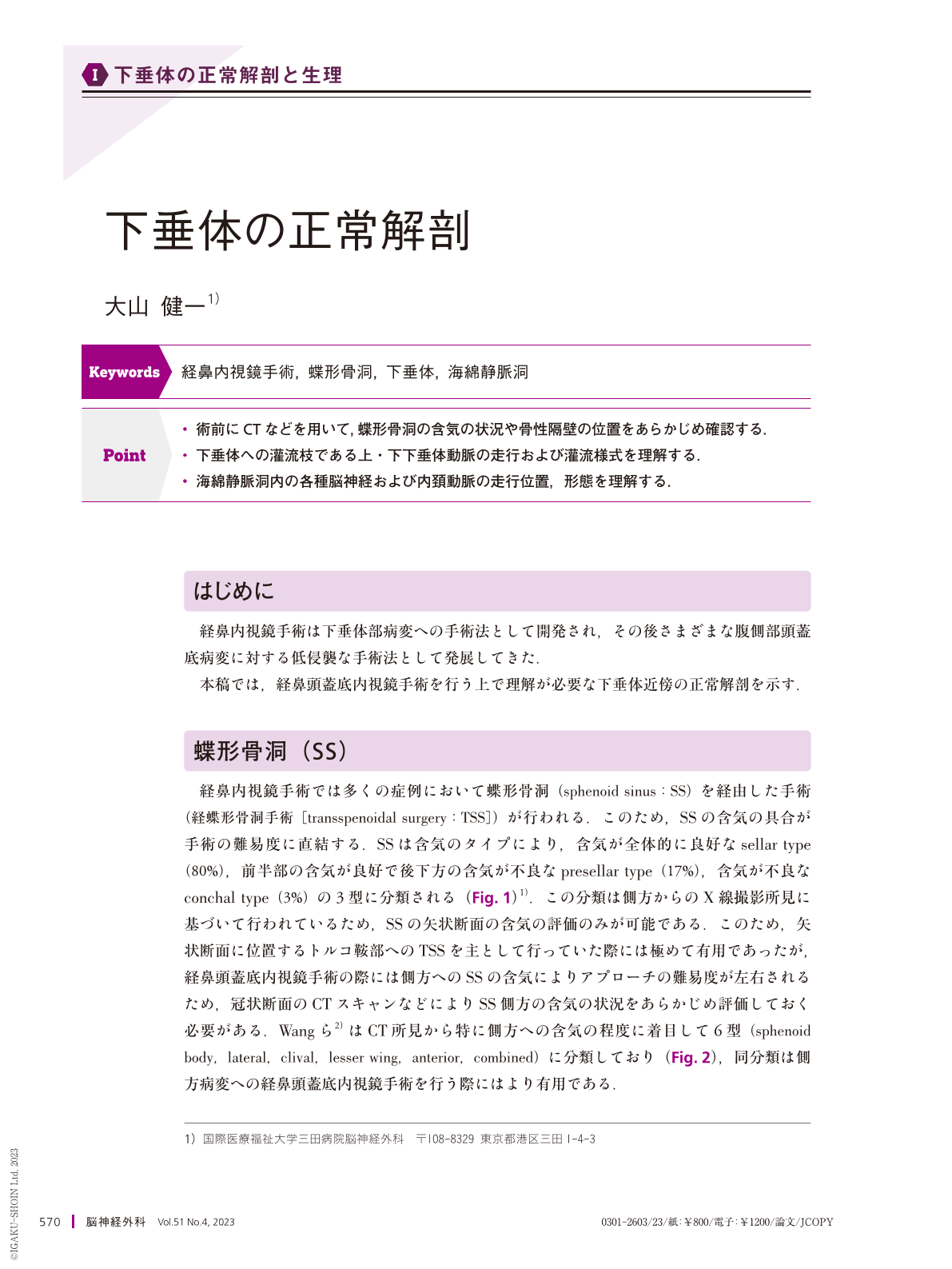
Point
・術前にCTなどを用いて,蝶形骨洞の含気の状況や骨性隔壁の位置をあらかじめ確認する.
・下垂体への灌流枝である上・下下垂体動脈の走行および灌流様式を理解する.
・海綿静脈洞内の各種脳神経および内頚動脈の走行位置,形態を理解する.
This study describes the anatomy of the pituitary gland during endoscopic endonasal surgery. Before surgery, the extent of pneumatization of the sphenoid sinus and bony septations in the sphenoid sinus should be evaluated using computed tomography. After wide sphenoidotomy, several important surgical landmarks, including the medial and lateral opticocarotid recesses and carotid protuberances, can be observed in the sphenoid sinus. The pituitary gland is composed of two components: the adenohypophysis and neurohypophysis. Two small vessels, the superior and inferior hypophyseal arteries, supply the pituitary gland. Several vital structures exist inside the cavernous sinus, including the internal carotid artery and cranial nerves.
Understanding the surgical anatomy is mandatory for treating lesions around the pituitary fossa via the endoscopic endonasal approach.
Copyright © 2023, Igaku-Shoin Ltd. All rights reserved.