Japanese
English
- 有料閲覧
PPV購入案内
1,320円 (1,200円+税10%) こちらは、801ページから808ページの文献です。購入には医書.jp本会員登録が必要です。
会員登録完了後に改めて上記「購入サイトへ」を選択してください。
または「購入サイトへ」選択後に「購入ログイン」、「新規会員登録」の順に進んでください。
会員登録について詳しくはをご確認ください。
- Abstract 文献概要
- 1ページ目 Look Inside
- 参考文献 Reference
Ⅰ.はじめに
Diffuse leptomeningeal glioneuronal tumor(DLGNT)は脳実質内への浸潤や腫瘍塊形成を認めずに,脳表の軟膜を中心にびまん性に分布することが特徴的な腫瘍である.過去の報告では,disseminated oligodendroglioma-like leptomeningeal neoplasmやprimary leptomeningeal oligodendro-gliomatosisなどの名称で呼ばれていたが,特徴的な病理組織学的・分子遺伝子学的性質を有することがわかり,World Health Organization(WHO)分類改訂第4版(2016年版)7)より独立した存在として記載された.小児から若年成人に多くみられ,やや男性に多い5,10,11,14).多くは緩徐な経過を呈するが,急性の経過を呈する例が一定数報告されている2,4,5,9-11,16).
今回われわれは,経過中にくも膜下出血を合併したDLGNTの1例を経験した.文献的考察を加えて報告する.
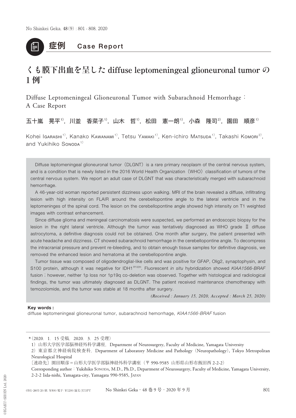
Diffuse leptomeningeal glioneuronal tumor(DLGNT)is a rare primary neoplasm of the central nervous system, and is a condition that is newly listed in the 2016 World Health Organization(WHO)classification of tumors of the central nervous system. We report an adult case of DLGNT that was characteristically merged with subarachnoid hemorrhage.
A 46-year-old woman reported persistent dizziness upon walking. MRI of the brain revealed a diffuse, infiltrating lesion with high intensity on FLAIR around the cerebellopontine angle to the lateral ventricle and in the leptomeninges of the spinal cord. The lesion on the cerebellopontine angle showed high intensity on T1 weighted images with contrast enhancement.
Since diffuse glioma and meningeal carcinomatosis were suspected, we performed an endoscopic biopsy for the lesion in the right lateral ventricle. Although the tumor was tentatively diagnosed as WHO grade Ⅱ diffuse astrocytoma, a definitive diagnosis could not be obtained. One month after surgery, the patient presented with acute headache and dizziness. CT showed subarachnoid hemorrhage in the cerebellopontine angle. To decompress the intracranial pressure and prevent re-bleeding, and to obtain enough tissue samples for definitive diagnosis, we removed the enhanced lesion and hematoma at the cerebellopontine angle.
Tumor tissue was composed of oligodendroglial-like cells and was positive for GFAP, Olig2, synaptophysin, and S100 protein, although it was negative for IDH1R132H. Fluorescent in situ hybridization showed KIAA1566-BRAF fusion;however, neither 1p loss nor 1p19q co-deletion was observed. Together with histological and radiological findings, the tumor was ultimately diagnosed as DLGNT. The patient received maintenance chemotherapy with temozolomide, and the tumor was stable at 18 months after surgery.
Copyright © 2020, Igaku-Shoin Ltd. All rights reserved.