Japanese
English
- 有料閲覧
PPV購入案内
1,320円 (1,200円+税10%) こちらは、321ページから327ページの文献です。購入には医書.jp本会員登録が必要です。
会員登録完了後に改めて上記「購入サイトへ」を選択してください。
または「購入サイトへ」選択後に「購入ログイン」、「新規会員登録」の順に進んでください。
会員登録について詳しくはをご確認ください。
- Abstract 文献概要
- 1ページ目 Look Inside
- 参考文献 Reference
Ⅰ.はじめに
脳動脈瘤のクリッピング術において,動脈瘤および親動脈の同定に必要かつ十分な術野を得るためには,術中早期から動脈瘤の位置を予測できることが望ましい.しかし,遠位部前大脳動脈瘤では,早期に動脈瘤の位置を正確に予測し,最短距離でアプローチすることが時に困難である.その理由として,①発生する部位や形状などが多岐にわたる,②指標となる構造物が少なく,かつ深部に存在する,③破裂脳動脈瘤が前頭葉に血腫を形成し,偏位することがある9,13),④遠位部前大脳動脈瘤の頻度が2.0〜6.7%と低く10),1人の術者が多くの手術を経験することは難しい,といったことが挙げられる.
そこでわれわれは,遠位部前大脳動脈瘤の手術において早期に動脈瘤の位置,親動脈との位置関係を把握するために,カラードップラーエコーを使用している.本稿ではその有用性について検討し報告する.
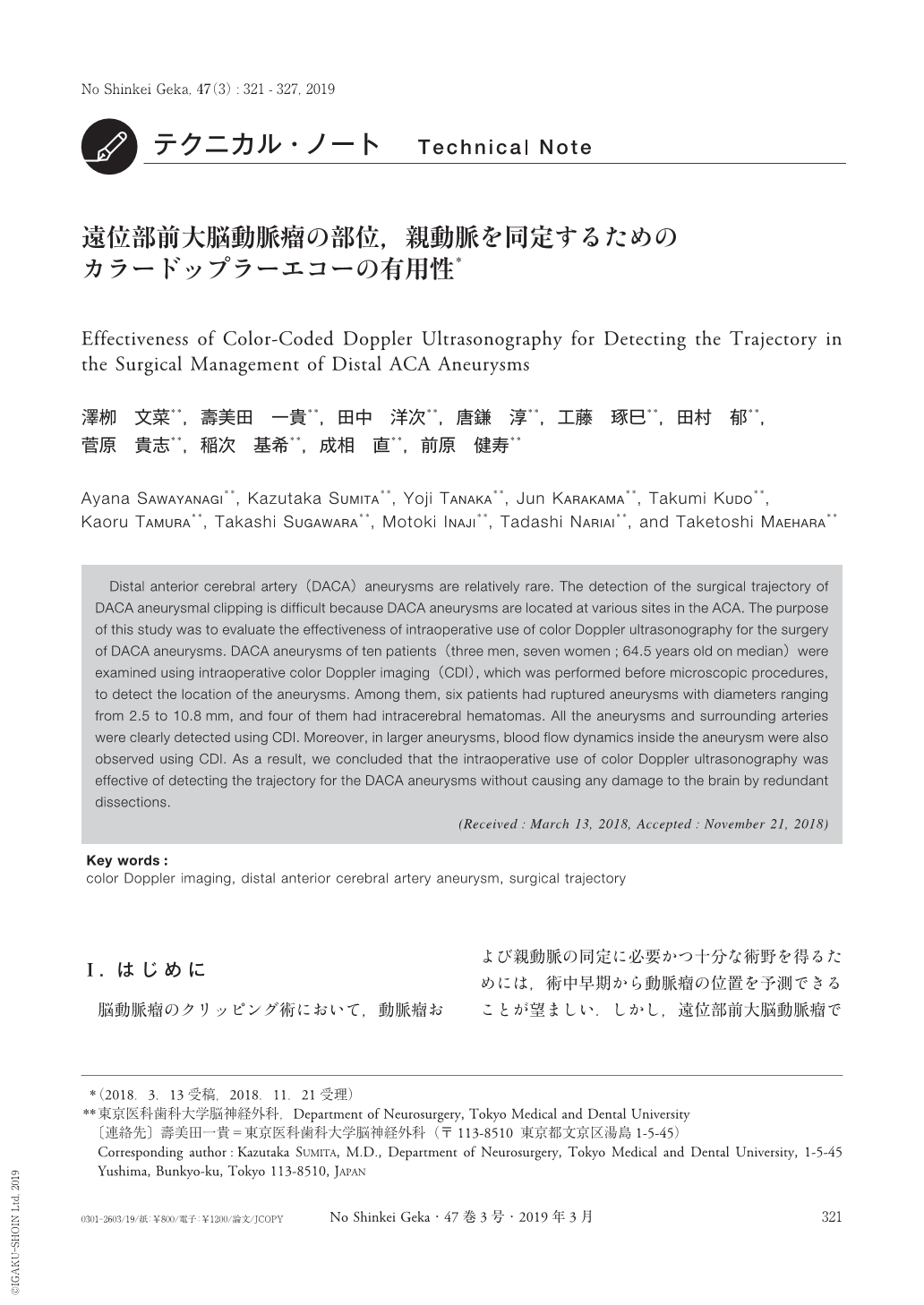
Distal anterior cerebral artery(DACA)aneurysms are relatively rare. The detection of the surgical trajectory of DACA aneurysmal clipping is difficult because DACA aneurysms are located at various sites in the ACA. The purpose of this study was to evaluate the effectiveness of intraoperative use of color Doppler ultrasonography for the surgery of DACA aneurysms. DACA aneurysms of ten patients(three men, seven women;64.5 years old on median)were examined using intraoperative color Doppler imaging(CDI), which was performed before microscopic procedures, to detect the location of the aneurysms. Among them, six patients had ruptured aneurysms with diameters ranging from 2.5 to 10.8mm, and four of them had intracerebral hematomas. All the aneurysms and surrounding arteries were clearly detected using CDI. Moreover, in larger aneurysms, blood flow dynamics inside the aneurysm were also observed using CDI. As a result, we concluded that the intraoperative use of color Doppler ultrasonography was effective of detecting the trajectory for the DACA aneurysms without causing any damage to the brain by redundant dissections.
Copyright © 2019, Igaku-Shoin Ltd. All rights reserved.