Japanese
English
- 有料閲覧
PPV購入案内
1,320円 (1,200円+税10%) こちらは、5ページから9ページの文献です。購入には医書.jp本会員登録が必要です。
会員登録完了後に改めて上記「購入サイトへ」を選択してください。
または「購入サイトへ」選択後に「購入ログイン」、「新規会員登録」の順に進んでください。
会員登録について詳しくはをご確認ください。
- Abstract 文献概要
- 1ページ目 Look Inside
- 参考文献 Reference
Ⅰ.はじめに
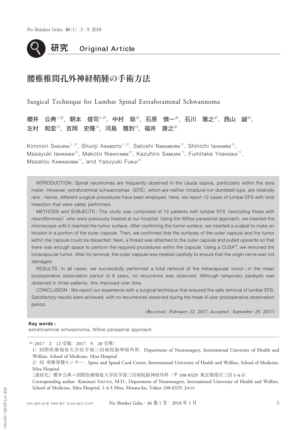
脊髄神経鞘腫は硬膜内,特に馬尾レベルに多いが,比較的大きいものであっても,顕微鏡下で手術を行えば安全に合併症なく全摘出可能である.しかし,硬膜外で,かつdumbbell typeではない神経鞘腫(extraforaminal schwannoma:EFS)は比較的珍しく,手術方法も大きく異なる.今回われわれは,腰椎EFSの摘出術を安全に行うための手術方法について,自験例をもとに検討した.
INTRODUCTION:Spinal neurinomas are frequently observed in the cauda equina, particularly within the dura mater. However, extraforaminal schwannomas(EFS), which are neither intradural nor dumbbell type, are relatively rare;hence, different surgical procedures have been employed. Here, we report 12 cases of lumbar EFS with total resection that were safely performed.
METHODS and SUBJECTS:This study was comprised of 12 patients with lumbar EFS(excluding those with neurofibromas)who were previously treated at our hospital. Using the Wiltse paraspinal approach, we inserted the microscope until it reached the tumor surface. After confirming the tumor surface, we inserted a scalpel to make an incision in a portion of the outer capsule. Then, we confirmed that the surfaces of the outer capsule and the tumor within the capsule could be dissected. Next, a thread was attached to the outer capsule and pulled upwards so that there was enough space to perform the required procedures within the capsule. Using a CUSA®, we removed the intracapsular tumor. After its removal, the outer capsule was treated carefully to ensure that the origin nerve was not damaged.
RESULTS:In all cases, we successfully performed a total removal of the intracapsular tumor;in the mean postoperative observation period of 8 years, no recurrence was observed. Although temporary paralysis was observed in three patients, this improved over time.
CONCLUSION:We report our experience with a surgical technique that ensured the safe removal of lumbar EFS. Satisfactory results were achieved, with no recurrences observed during the mean 8-year postoperative observation period.
Copyright © 2018, Igaku-Shoin Ltd. All rights reserved.