Japanese
English
- 有料閲覧
PPV購入案内
1,320円 (1,200円+税10%) こちらは、1001ページから1009ページの文献です。購入には医書.jp本会員登録が必要です。
会員登録完了後に改めて上記「購入サイトへ」を選択してください。
または「購入サイトへ」選択後に「購入ログイン」、「新規会員登録」の順に進んでください。
会員登録について詳しくはをご確認ください。
- Abstract 文献概要
- 1ページ目 Look Inside
- 参考文献 Reference
Ⅰ.はじめに
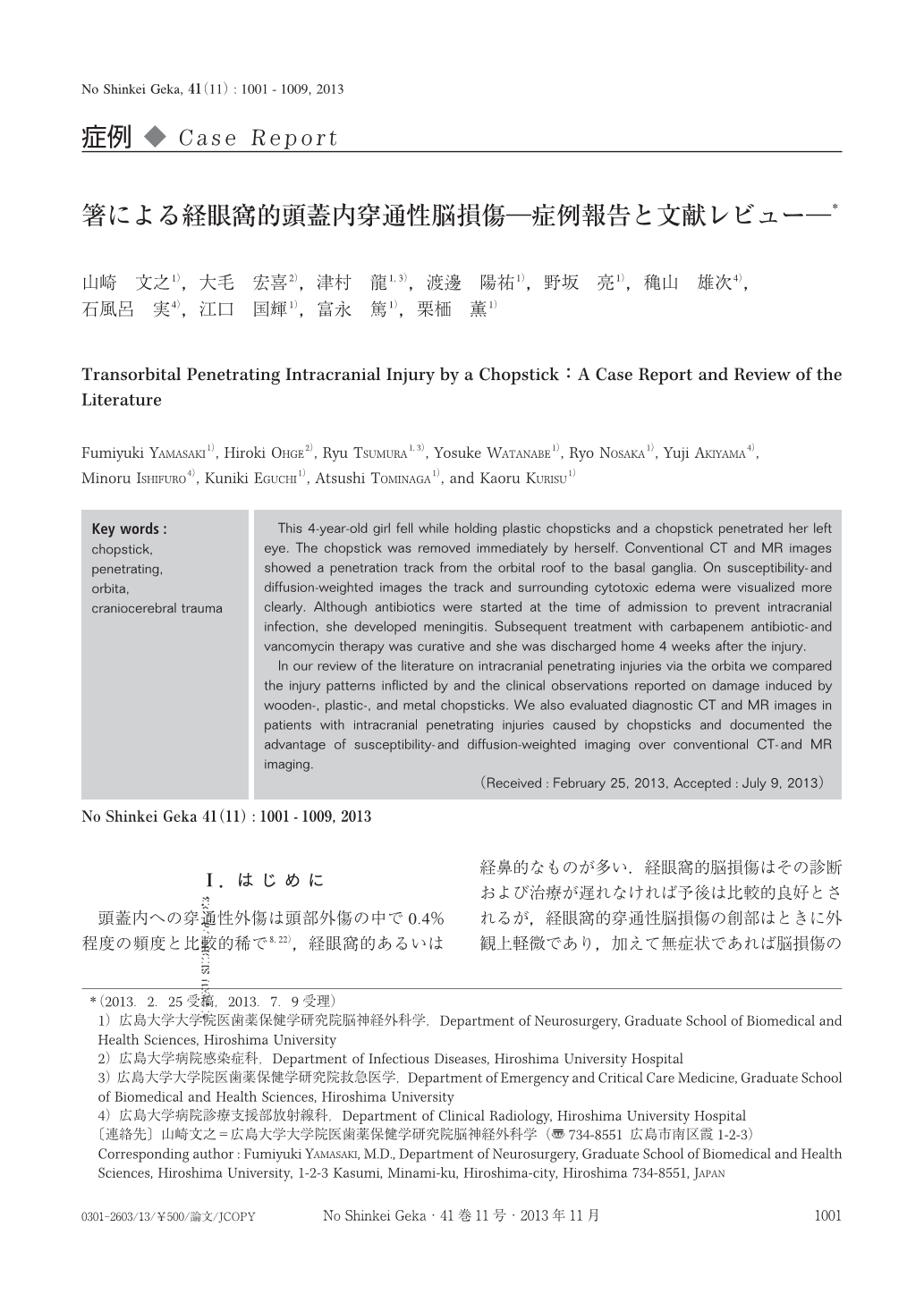
頭蓋内への穿通性外傷は頭部外傷の中で0.4%程度の頻度と比較的稀で8,22),経眼窩的あるいは経鼻的なものが多い.経眼窩的脳損傷はその診断および治療が遅れなければ予後は比較的良好とされるが,経眼窩的穿通性脳損傷の創部はときに外観上軽微であり,加えて無症状であれば脳損傷の診断は困難なことがある.今回われわれは箸による経眼窩的頭蓋内穿通性脳損傷の1例を経験したので,その画像診断の有用性や問題点を文献的に考察して報告する.また,箸による経眼窩的頭蓋内穿通性脳損傷の特徴を文献的に検討する.
This 4-year-old girl fell while holding plastic chopsticks and a chopstick penetrated her left eye. The chopstick was removed immediately by herself. Conventional CT and MR images showed a penetration track from the orbital roof to the basal ganglia. On susceptibility-and diffusion-weighted images the track and surrounding cytotoxic edema were visualized more clearly. Although antibiotics were started at the time of admission to prevent intracranial infection, she developed meningitis. Subsequent treatment with carbapenem antibiotic-and vancomycin therapy was curative and she was discharged home 4 weeks after the injury.
In our review of the literature on intracranial penetrating injuries via the orbita we compared the injury patterns inflicted by and the clinical observations reported on damage induced by wooden-, plastic-, and metal chopsticks. We also evaluated diagnostic CT and MR images in patients with intracranial penetrating injuries caused by chopsticks and documented the advantage of susceptibility-and diffusion-weighted imaging over conventional CT-and MR imaging.
Copyright © 2013, Igaku-Shoin Ltd. All rights reserved.