Japanese
English
- 有料閲覧
- Abstract 文献概要
- 1ページ目 Look Inside
I.はじめに
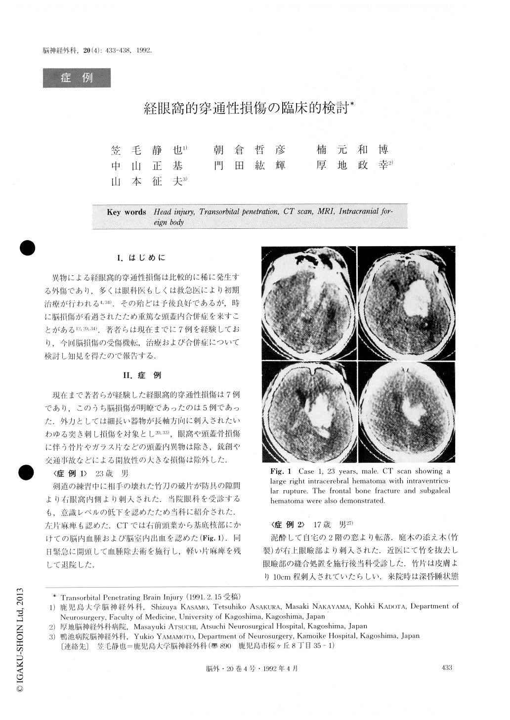
異物による経眼窩的穿通性損傷は比較的に稀に発生する外傷であり,多くは眼科医もしくは救急医により初期治療が行われる4,34).その殆どは予後良好であるが.時に脳損傷が看過されたため重篤な頭蓋内合併症を来すことがある12,20,34),著者らは現在までに7例を経験しており,今回脳損傷の受傷機転,治療および合併症について検討し知見を得たので報告する.
Transorbital penetrating brain injury is rare during this time of peace. In our paper, we reported sever cases of these injuries and discussed the mechanism and treatment of intracranial complications.
Transorbital penetrating brain injuries were caused by thin, long and relative hard objects such as chop-stick (case 3), pencil (case 6), bamboo stick (case 1, 2, 7) and a piece of metal (case 4, 5). CT scan, MRI and angiography demonstrated a large variety of intracra-nial complications. For instance. intracerebral hemato-ma, cerebral contusion, intraventricular hemorrhage, pneumocephalus, brain stem injury and carotid caver-nous sinus fistula. We had no case of infectious com-plications such as meningitis and brain abscess. If the direction of the injuring object runs parallel to the orbital roof, it penetrates the cranial cavity com-monly via the superior orbital fissure or the optic canal, which routes provide direct access without hone frac-ture. This direction will cause critical intracranial com-plications such as CCF or brain stem injury. If the in-juring object runs upward and across the orbital roof which has thin bone and therefore offers little resis-tance, the frontal lobe will be easily damaged, and it will cause intracerebral hematoma. But the prognosis for this type of injury is not so poor because we can easily remove the hematoma and the foreign body. With our treatment policy of "pull and see, all our cases but one recovered well. The one exception was a case in which a large intracerebral hematoma was over-looked in an ophthalomological clinic. This patient died. Nowadays, CT scan and MRI give clear informa-tion about the anatomical location of injuring objects and intracranial complications. Instruments and mate-rials for use in neurosurgical operations, and neurosur-gical techniques have advanced more and more. So we think that early diagnosis and early treatment including craniotomy are the best therapies for transorbital pene-trating brain injury.
Copyright © 1992, Igaku-Shoin Ltd. All rights reserved.