Japanese
English
- 有料閲覧
PPV購入案内
1,320円 (1,200円+税10%) こちらは、643ページから650ページの文献です。購入には医書.jp本会員登録が必要です。
会員登録完了後に改めて上記「購入サイトへ」を選択してください。
または「購入サイトへ」選択後に「購入ログイン」、「新規会員登録」の順に進んでください。
会員登録について詳しくはをご確認ください。
- Abstract 文献概要
- 1ページ目 Look Inside
- 参考文献 Reference
Ⅰ.はじめに
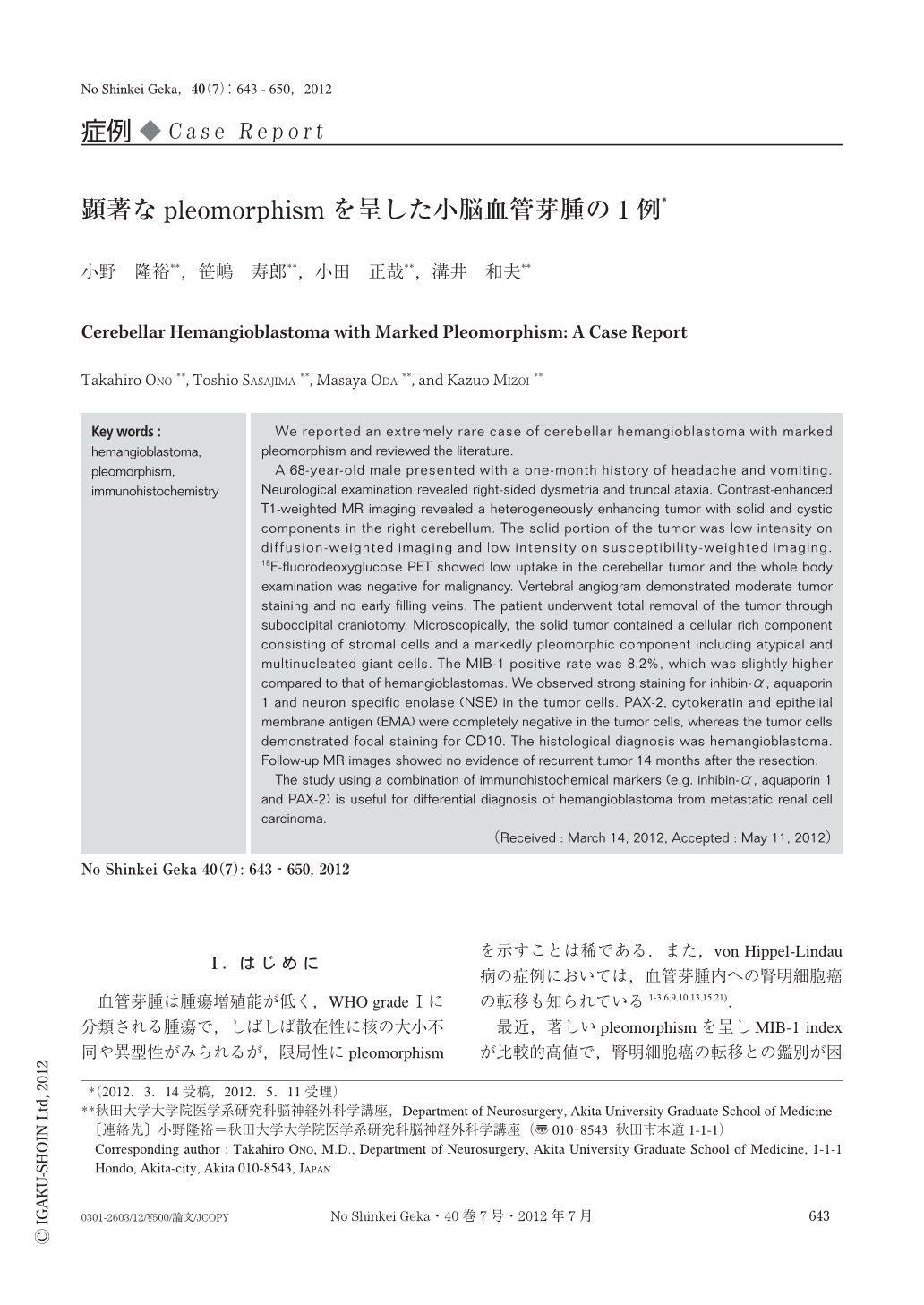
血管芽腫は腫瘍増殖能が低く,WHO gradeⅠに分類される腫瘍で,しばしば散在性に核の大小不同や異型性がみられるが,限局性にpleomorphismを示すことは稀である.また,von Hippel-Lindau病の症例においては,血管芽腫内への腎明細胞癌の転移も知られている1-3,6,9,10,13,15,21).
最近,著しいpleomorphismを呈しMIB-1 indexが比較的高値で,腎明細胞癌の転移との鑑別が困難であった小脳血管芽腫の1例を経験したので,文献的考察を加えて報告する.
We reported an extremely rare case of cerebellar hemangioblastoma with marked pleomorphism and reviewed the literature.
A 68-year-old male presented with a one-month history of headache and vomiting. Neurological examination revealed right-sided dysmetria and truncal ataxia. Contrast-enhanced T1-weighted MR imaging revealed a heterogeneously enhancing tumor with solid and cystic components in the right cerebellum. The solid portion of the tumor was low intensity on diffusion-weighted imaging and low intensity on susceptibility-weighted imaging. 18F-fluorodeoxyglucose PET showed low uptake in the cerebellar tumor and the whole body examination was negative for malignancy. Vertebral angiogram demonstrated moderate tumor staining and no early filling veins. The patient underwent total removal of the tumor through suboccipital craniotomy. Microscopically, the solid tumor contained a cellular rich component consisting of stromal cells and a markedly pleomorphic component including atypical and multinucleated giant cells. The MIB-1 positive rate was 8.2%, which was slightly higher compared to that of hemangioblastomas. We observed strong staining for inhibin-α, aquaporin 1 and neuron specific enolase (NSE) in the tumor cells. PAX-2, cytokeratin and epithelial membrane antigen (EMA) were completely negative in the tumor cells, whereas the tumor cells demonstrated focal staining for CD10. The histological diagnosis was hemangioblastoma. Follow-up MR images showed no evidence of recurrent tumor 14 months after the resection.
The study using a combination of immunohistochemical markers (e.g. inhibin-α,aquaporin 1 and PAX-2) is useful for differential diagnosis of hemangioblastoma from metastatic renal cell carcinoma.
Copyright © 2012, Igaku-Shoin Ltd. All rights reserved.