Japanese
English
- 有料閲覧
PPV購入案内
1,320円 (1,200円+税10%) こちらは、623ページから628ページの文献です。購入には医書.jp本会員登録が必要です。
会員登録完了後に改めて上記「購入サイトへ」を選択してください。
または「購入サイトへ」選択後に「購入ログイン」、「新規会員登録」の順に進んでください。
会員登録について詳しくはをご確認ください。
- Abstract 文献概要
- 1ページ目 Look Inside
- 参考文献 Reference
Ⅰ.はじめに
孤立性骨囊腫(solitary bone cyst:SBC)は骨の類腫瘍性疾患に分類され,大腿骨や上腕骨など長管骨に好発し,頭蓋骨発生例は稀である3-6,9-13).今回,打撲を契機に腫瘤の増大傾向を示したと推察できる頭蓋骨発生のSBCを経験したので,文献的考察を加えて報告する.
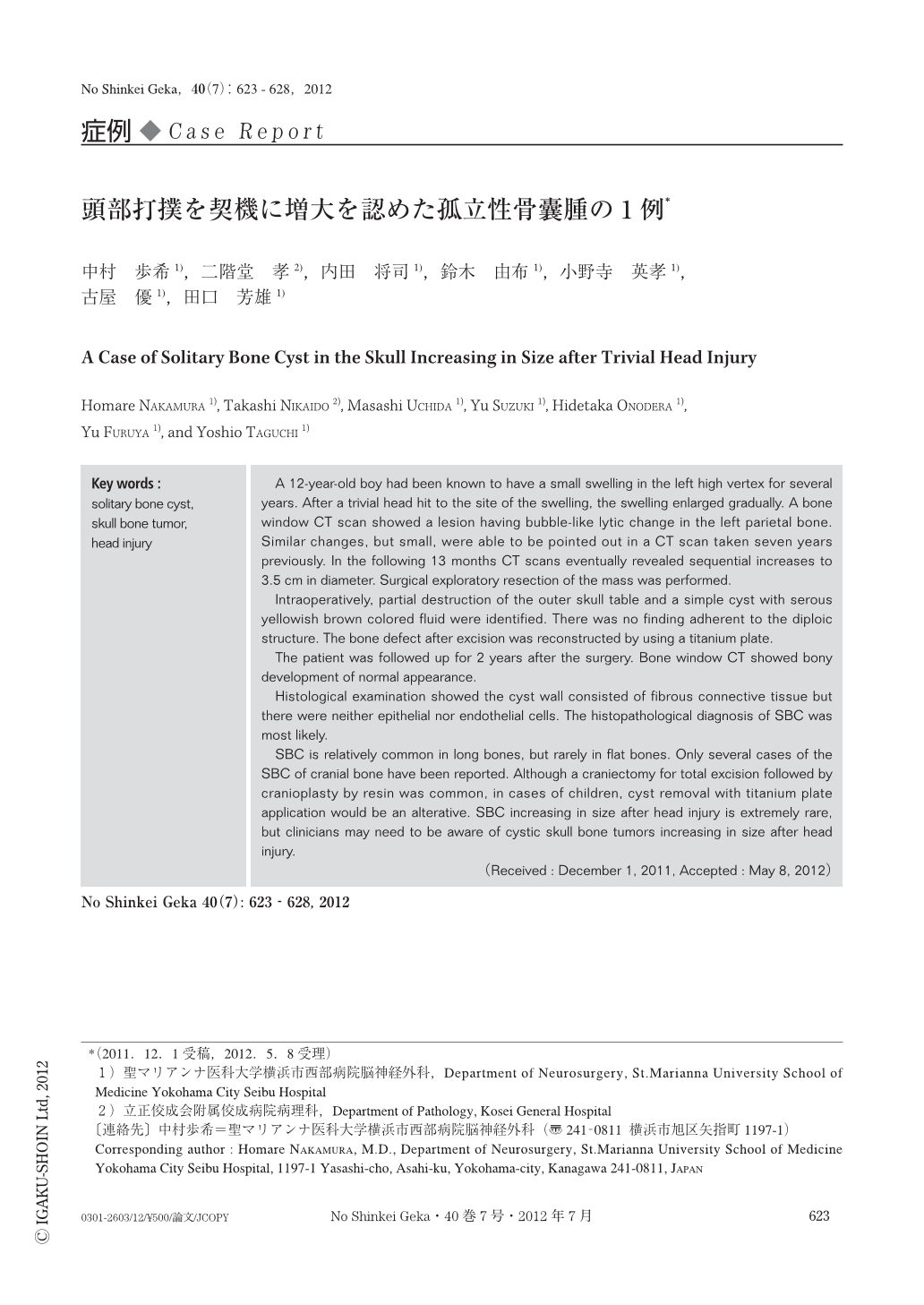
A 12-year-old boy had been known to have a small swelling in the left high vertex for several years. After a trivial head hit to the site of the swelling,the swelling enlarged gradually. A bone window CT scan showed a lesion having bubble-like lytic change in the left parietal bone. Similar changes,but small,were able to be pointed out in a CT scan taken seven years previously. In the following 13 months CT scans eventually revealed sequential increases to 3.5 cm in diameter. Surgical exploratory resection of the mass was performed.
Intraoperatively, partial destruction of the outer skull table and a simple cyst with serous yellowish brown colored fluid were identified. There was no finding adherent to the diploic structure. The bone defect after excision was reconstructed by using a titanium plate.
The patient was followed up for 2 years after the surgery. Bone window CT showed bony development of normal appearance.
Histological examination showed the cyst wall consisted of fibrous connective tissue but there were neither epithelial nor endothelial cells. The histopathological diagnosis of SBC was most likely.
SBC is relatively common in long bones,but rarely in flat bones. Only several cases of the SBC of cranial bone have been reported. Although a craniectomy for total excision followed by cranioplasty by resin was common,in cases of children,cyst removal with titanium plate application would be an alterative. SBC increasing in size after head injury is extremely rare,but clinicians may need to be aware of cystic skull bone tumors increasing in size after head injury.
Copyright © 2012, Igaku-Shoin Ltd. All rights reserved.