Japanese
English
- 有料閲覧
PPV購入案内
1,320円 (1,200円+税10%) こちらは、157ページから162ページの文献です。購入には医書.jp本会員登録が必要です。
会員登録完了後に改めて上記「購入サイトへ」を選択してください。
または「購入サイトへ」選択後に「購入ログイン」、「新規会員登録」の順に進んでください。
会員登録について詳しくはをご確認ください。
- Abstract 文献概要
- 1ページ目 Look Inside
- 参考文献 Reference
Ⅰ.はじめに
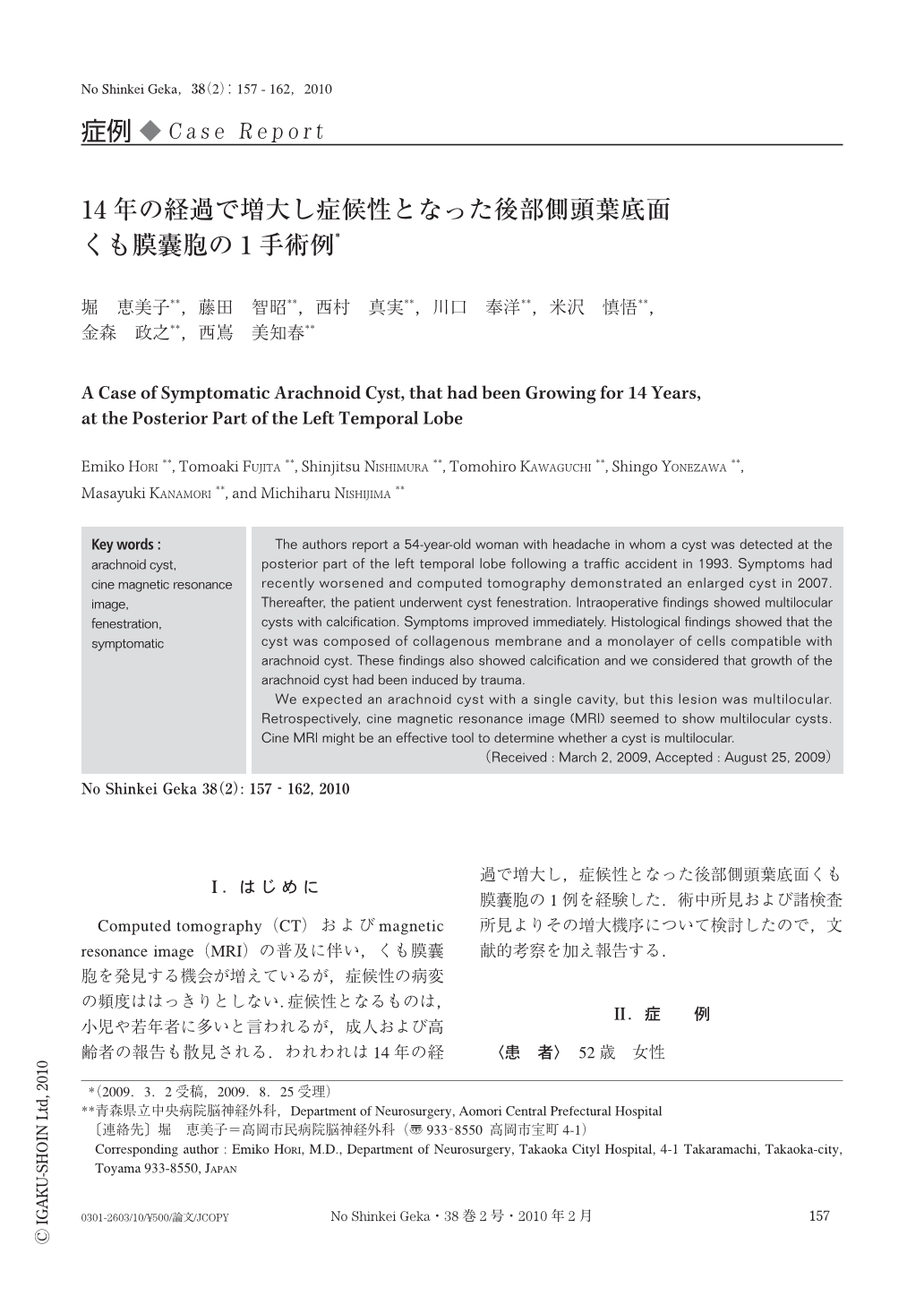
Computed tomography(CT)およびmagnetic resonance image(MRI)の普及に伴い,くも膜囊胞を発見する機会が増えているが,症候性の病変の頻度ははっきりとしない.症候性となるものは,小児や若年者に多いと言われるが,成人および高齢者の報告も散見される.われわれは14年の経過で増大し,症候性となった後部側頭葉底面くも膜囊胞の1例を経験した.術中所見および諸検査所見よりその増大機序について検討したので,文献的考察を加え報告する.
The authors report a 54-year-old woman with headache in whom a cyst was detected at the posterior part of the left temporal lobe following a traffic accident in 1993. Symptoms had recently worsened and computed tomography demonstrated an enlarged cyst in 2007. Thereafter, the patient underwent cyst fenestration. Intraoperative findings showed multilocular cysts with calcification. Symptoms improved immediately. Histological findings showed that the cyst was composed of collagenous membrane and a monolayer of cells compatible with arachnoid cyst. These findings also showed calcification and we considered that growth of the arachnoid cyst had been induced by trauma.
We expected an arachnoid cyst with a single cavity, but this lesion was multilocular. Retrospectively, cine magnetic resonance image (MRI) seemed to show multilocular cysts. Cine MRI might be an effective tool to determine whether a cyst is multilocular.
Copyright © 2010, Igaku-Shoin Ltd. All rights reserved.