Japanese
English
- 有料閲覧
PPV購入案内
1,320円 (1,200円+税10%) こちらは、167ページから171ページの文献です。購入には医書.jp本会員登録が必要です。
会員登録完了後に改めて上記「購入サイトへ」を選択してください。
または「購入サイトへ」選択後に「購入ログイン」、「新規会員登録」の順に進んでください。
会員登録について詳しくはをご確認ください。
- Abstract 文献概要
- 1ページ目 Look Inside
- 参考文献 Reference
Ⅰ.はじめに
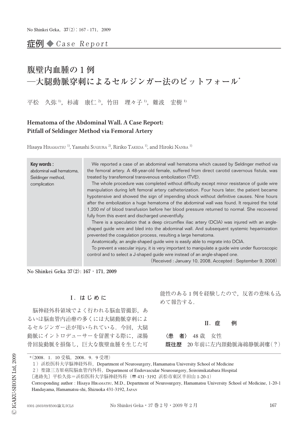
脳神経外科領域でよく行われる脳血管撮影,あるいは脳血管内治療の多くには大腿動脈穿刺によるセルジンガー法が用いられている.今回,大腿動脈にイントロデューサーを留置する際に,深腸骨回旋動脈を損傷し,巨大な腹壁血腫を生じた可能性のある1例を経験したので,反省の意味も込めて報告する.
We reported a case of an abdominal wall hematoma which caused by Seldinger method via the femoral artery. A 48-year-old female, suffered from direct carotid cavernous fistula, was treated by transfemoral transvenous embolization (TVE).
The whole procedure was completed without difficulty except minor resistance of guide wire manipulation during left femoral artery catheterization. Four hours later, the patient became hypotensive and showed the sign of impending shock without definitive causes. Nine hours after the embolization a huge hematoma of the abdominal wall was found. It required the total 1,200ml of blood transfusion before her blood pressure returned to normal. She recovered fully from this event and discharged uneventfully.
There is a speculation that a deep circumflex iliac artery (DCIA) was injured with an angle-shaped guide wire and bled into the abdominal wall. And subsequent systemic heparinization prevented the coagulation process, resulting a large hematoma.
Anatomically, an angle-shaped guide wire is easily able to migrate into DCIA.
To prevent a vascular injury, it is very important to manipulate a guide wire under fluoroscopic control and to select a J-shaped guide wire instead of an angle-shaped one.
Copyright © 2009, Igaku-Shoin Ltd. All rights reserved.