Japanese
English
- 有料閲覧
PPV購入案内
1,320円 (1,200円+税10%) こちらは、955ページから959ページの文献です。購入には医書.jp本会員登録が必要です。
会員登録完了後に改めて上記「購入サイトへ」を選択してください。
または「購入サイトへ」選択後に「購入ログイン」、「新規会員登録」の順に進んでください。
会員登録について詳しくはをご確認ください。
- Abstract 文献概要
- 1ページ目 Look Inside
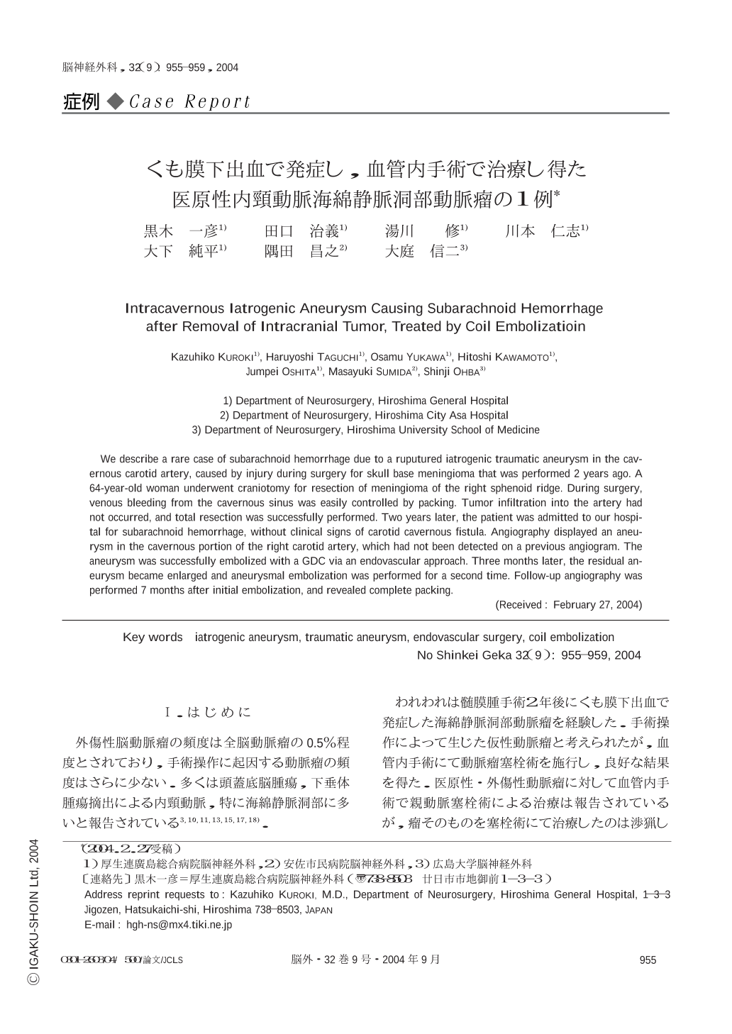
Ⅰ.はじめに
外傷性脳動脈瘤の頻度は全脳動脈瘤の0.5%程度とされており,手術操作に起因する動脈瘤の頻度はさらに少ない.多くは頭蓋底脳腫瘍,下垂体腫瘍摘出による内頸動脈,特に海綿静脈洞部に多いと報告されている3,10,11,13,15,17,18).
われわれは髄膜腫手術2年後にくも膜下出血で発症した海綿静脈洞部動脈瘤を経験した.手術操作によって生じた仮性動脈瘤と考えられたが,血管内手術にて動脈瘤塞栓術を施行し,良好な結果を得た.医原性・外傷性動脈瘤に対して血管内手術で親動脈塞栓術による治療は報告されているが,瘤そのものを塞栓術にて治療したのは渉猟し得た限りでは5例しかない.病態・治療を文献的考察を加え,報告する.
We describe a rare case of subarachnoid hemorrhage due to a ruputured iatrogenic traumatic aneurysm in the cavernous carotid artery,caused by injury during surgery for skull base meningioma that was performed 2 years ago. A 64-year-old woman underwent craniotomy for resection of meningioma of the right sphenoid ridge. During surgery,venous bleeding from the cavernous sinus was easily controlled by packing. Tumor infiltration into the artery had not occurred,and total resection was successfully performed. Two years later,the patient was admitted to our hospital for subarachnoid hemorrhage,without clinical signs of carotid cavernous fistula. Angiography displayed an aneurysm in the cavernous portion of the right carotid artery,which had not been detected on a previous angiogram. The aneurysm was successfully embolized with a GDC via an endovascular approach. Three months later,the residual aneurysm became enlarged and aneurysmal embolization was performed for a second time. Follow-up angiography was performed 7 months after initial embolization,and revealed complete packing.
Copyright © 2004, Igaku-Shoin Ltd. All rights reserved.