Japanese
English
- 有料閲覧
PPV購入案内
1,320円 (1,200円+税10%) こちらは、261ページから266ページの文献です。購入には医書.jp本会員登録が必要です。
会員登録完了後に改めて上記「購入サイトへ」を選択してください。
または「購入サイトへ」選択後に「購入ログイン」、「新規会員登録」の順に進んでください。
会員登録について詳しくはをご確認ください。
- Abstract 文献概要
- 1ページ目 Look Inside
I.はじめに
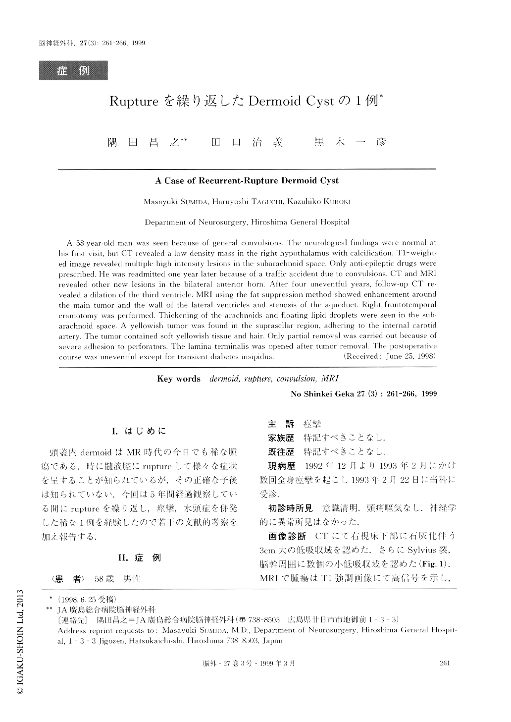
頭蓋内dermoidはMR時代の今日でも稀な腫瘍である.時に髄液腔にruptureして様々な症状を呈することが知られているが,その正確な予後は知られていない.今回は5年間経過観察している間にruptureを繰り返し,痙攣,水頭症を併発した稀な1例を経験したので若干の文献的考察を加え報告する.
A 58-year-old man was seen because of general convulsions. The neurological findings were normal athis first visit, but CT revealed a low density mass in the right hypothalamus with calcification. T1-weight-ed image revealed multiple high intensity lesions in the subarachnoid space. Only anti-epileptic drugs wereprescribed. He was readmitted one year later because of a traffic accident due to convulsions. CT and MRIrevealed other new lesions in the bilateral anterior horn. After four uneventful years, follow-up CT re-vealed a dilation of the third ventricle. MRI using the fat suppression method showed enhancement aroundthe main tumor and the wall of the lateral ventricles and stenosis of the aqueduct. Right frontotemporalcraniotomy was performed. Thickening of the arachnoids and floating lipid droplets were seen in the sub-arachnoid space. A yellowish tumor was found in the suprasellar region, adhering to the internal carotidartery. The tumor contained soft yellowish tissueand hair. Only partial removal was carried out because ofsevere adhesion to perforators. The lamina terminalis was opened after tumor removal. The postoperativecourse was uneventful except for transient diabetes insipidus.
Copyright © 1999, Igaku-Shoin Ltd. All rights reserved.