Japanese
English
- 有料閲覧
PPV購入案内
1,320円 (1,200円+税10%) こちらは、723ページから727ページの文献です。購入には医書.jp本会員登録が必要です。
会員登録完了後に改めて上記「購入サイトへ」を選択してください。
または「購入サイトへ」選択後に「購入ログイン」、「新規会員登録」の順に進んでください。
会員登録について詳しくはをご確認ください。
- Abstract 文献概要
- 1ページ目 Look Inside
- 参考文献 Reference
Ⅰ.は じ め に
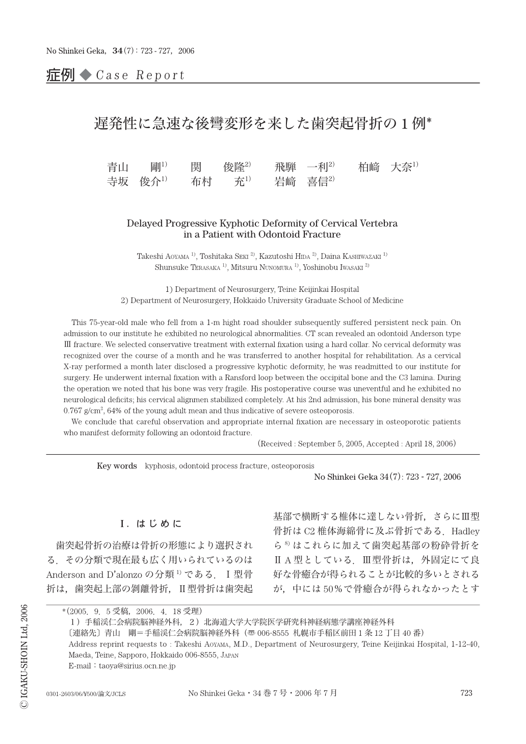
歯突起骨折の治療は骨折の形態により選択される.その分類で現在最も広く用いられているのはAnderson and D'alonzoの分類1)である.Ⅰ型骨折は,歯突起上部の剝離骨折,Ⅱ型骨折は歯突起基部で横断する椎体に達しない骨折,さらにⅢ型骨折はC2椎体海綿骨に及ぶ骨折である.Hadleyら8)はこれらに加えて歯突起基部の粉砕骨折をⅡA型としている.Ⅲ型骨折は,外固定にて良好な骨癒合が得られることが比較的多いとされるが,中には50%で骨癒合が得られなかったとする報告もあり3),治療上議論の多いところである.今回,硬性カラーによる外固定開始1カ月間は良好なアライメントが保持できたが,その後急速に後彎変形が進行し,内固定が必要となった症例の経験を報告する.
This 75-year-old male who fell from a 1-m hight road shoulder subsequently suffered persistent neck pain. On admission to our institute he exhibited no neurological abnormalities. CT scan revealed an odontoid Anderson type Ⅲ fracture. We selected conservative treatment with external fixation using a hard collar. No cervical deformity was recognized over the course of a month and he was transferred to another hospital for rehabilitation. As a cervical X-ray performed a month later disclosed a progressive kyphotic deformity,he was readmitted to our institute for surgery. He underwent internal fixation with a Ransford loop between the occipital bone and the C3 lamina. During the operation we noted that his bone was very fragile. His postoperative course was uneventful and he exhibited no neurological deficits; his cervical alignmen stabilized completely. At his 2nd admission,his bone mineral density was 0.767g/cm2,64% of the young adult mean and thus indicative of severe osteoporosis.
We conclude that careful observation and appropriate internal fixation are necessary in osteoporotic patients who manifest deformity following an odontoid fracture.
Copyright © 2006, Igaku-Shoin Ltd. All rights reserved.