Japanese
English
- 有料閲覧
- Abstract 文献概要
- 1ページ目 Look Inside
Ⅰ.はじめに
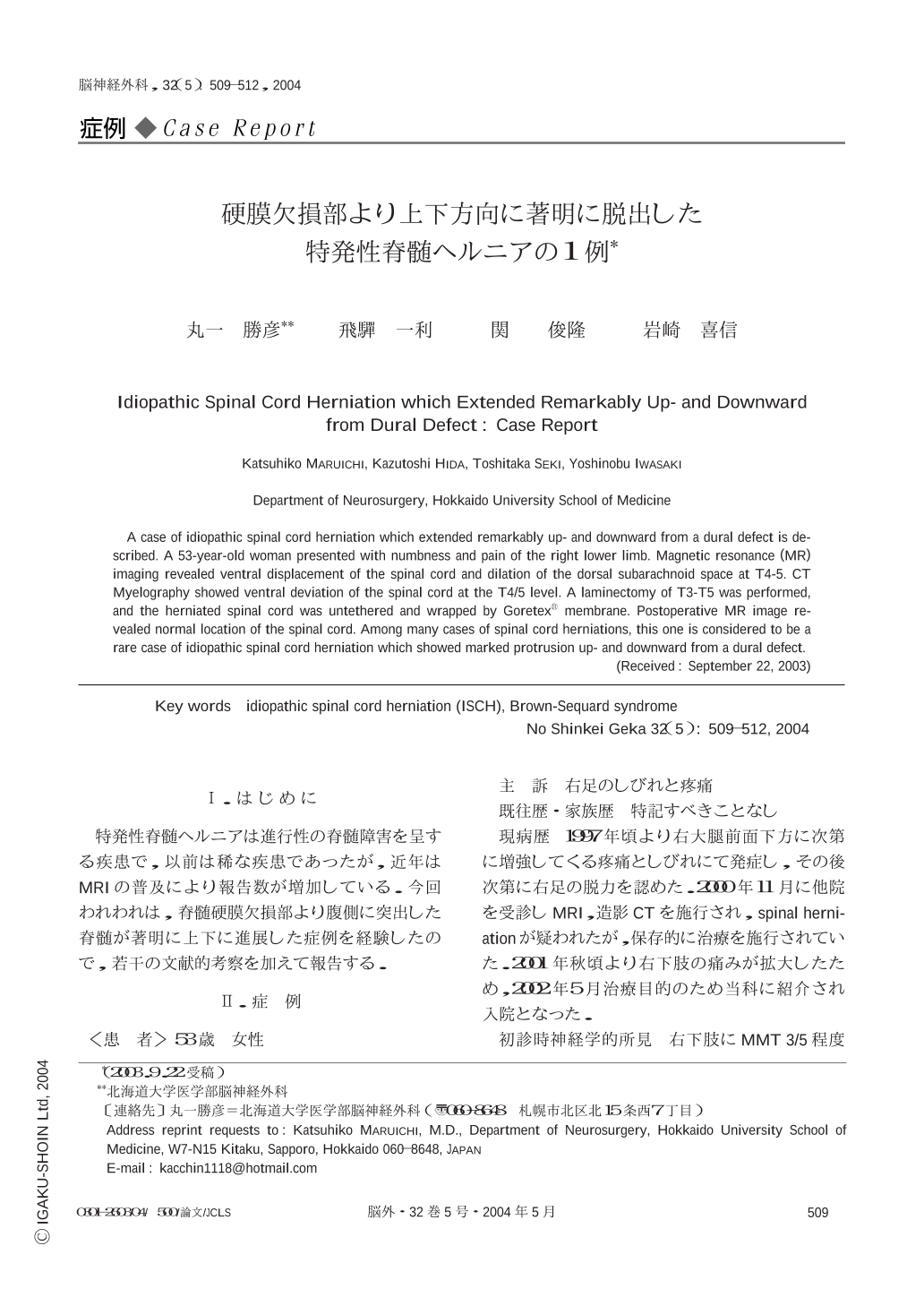
特発性脊髄ヘルニアは進行性の脊髄障害を呈する疾患で,以前は稀な疾患であったが,近年はMRIの普及により報告数が増加している.今回われわれは,脊髄硬膜欠損部より腹側に突出した脊髄が著明に上下に進展した症例を経験したので,若干の文献的考察を加えて報告する.
A case of idiopathic spinal cord herniation which extended remarkably up- and downward from a dural defect is described. A 53-year-old woman presented with numbness and pain of the right lower limb. Magnetic resonance (MR) imaging revealed ventral displacement of the spinal cord and dilation of the dorsal subarachnoid space at T4-5. CT Myelography showed ventral deviation of the spinal cord at the T4/5 level. A laminectomy of T3-T5 was performed,and the herniated spinal cord was untethered and wrapped by Goretex(R) membrane. Postoperative MR image revealed normal location of the spinal cord. Among many cases of spinal cord herniations,this one is considered to be a rare case of idiopathic spinal cord herniation which showed marked protrusion up- and downward from a dural defect.

Copyright © 2004, Igaku-Shoin Ltd. All rights reserved.


