Japanese
English
- 有料閲覧
PPV購入案内
1,320円 (1,200円+税10%) こちらは、629ページから634ページの文献です。購入には医書.jp本会員登録が必要です。
会員登録完了後に改めて上記「購入サイトへ」を選択してください。
または「購入サイトへ」選択後に「購入ログイン」、「新規会員登録」の順に進んでください。
会員登録について詳しくはをご確認ください。
- Abstract 文献概要
- 1ページ目 Look Inside
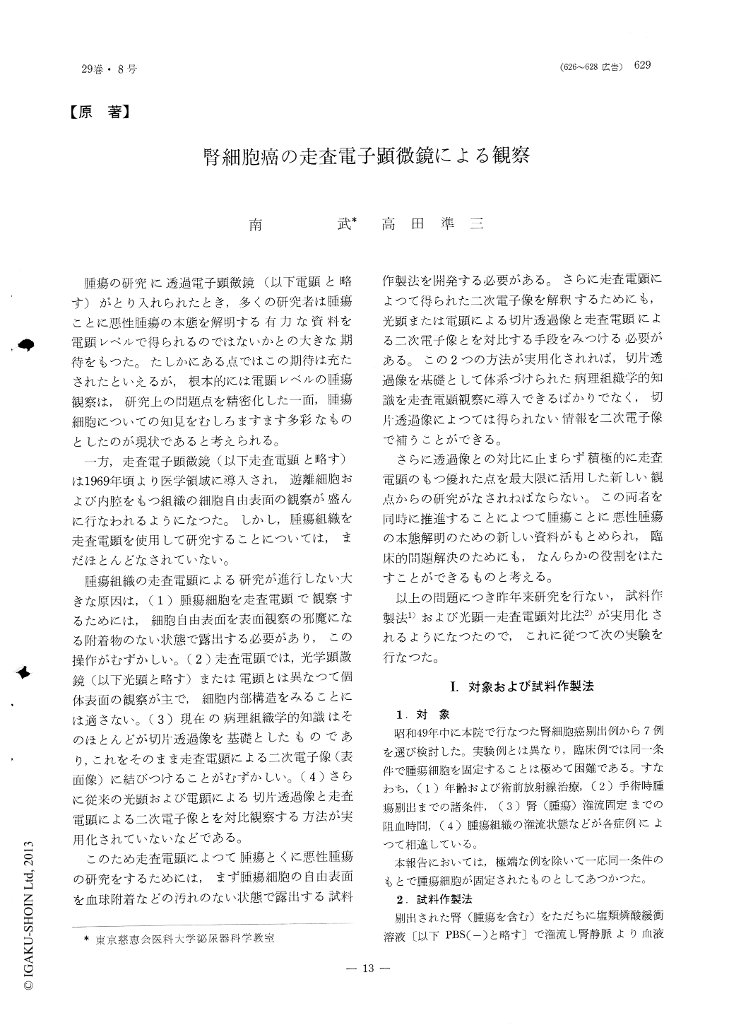
腫瘍の研究に透過電子顕微鏡(以下電顕と略す)がとり入れられたとき,多くの研究者は腫瘍ことに悪性腫瘍の本態を解明する有力な資料を電顕レベルで得られるのではないかとの大きな期待をもつた。たしかにある点ではこの期待は充たされたといえるが,根本的には電顕レベルの腫瘍観察は,研究上の問題点を精密化した一面,腫瘍細胞についての知見をむしろますます多彩なものとしたのが現状であると考えられる。
一方,走査電子顕微鏡(以下走査電顕と略す)は1969年頃より医学領域に導入され,遊離細胞および内腔をもつ組織の細胞自由表面の観察が盛んに行なわれるようになつた。しかし,腫瘍組織を走査電顕を使用して研究することについては,まだほとんどなされていない。
Studies of malignant tumors by scanning electron microscope at the present time, are limited to the observation of the surface of cultivated cancer cells. This is based on the fact that there are no adequate method to frame specimens for the observation of tumor tissue.
A method adequate to frame specimens for observation of the surface of cancer cells was designed and comparison of findings by scanning etectron microscope and optic microscope were done on 7 cases of renal cell carcinoma.
Copyright © 1975, Igaku-Shoin Ltd. All rights reserved.