Japanese
English
- 有料閲覧
PPV購入案内
1,320円 (1,200円+税10%) こちらは、505ページから510ページの文献です。購入には医書.jp本会員登録が必要です。
会員登録完了後に改めて上記「購入サイトへ」を選択してください。
または「購入サイトへ」選択後に「購入ログイン」、「新規会員登録」の順に進んでください。
会員登録について詳しくはをご確認ください。
- Abstract 文献概要
- 1ページ目 Look Inside
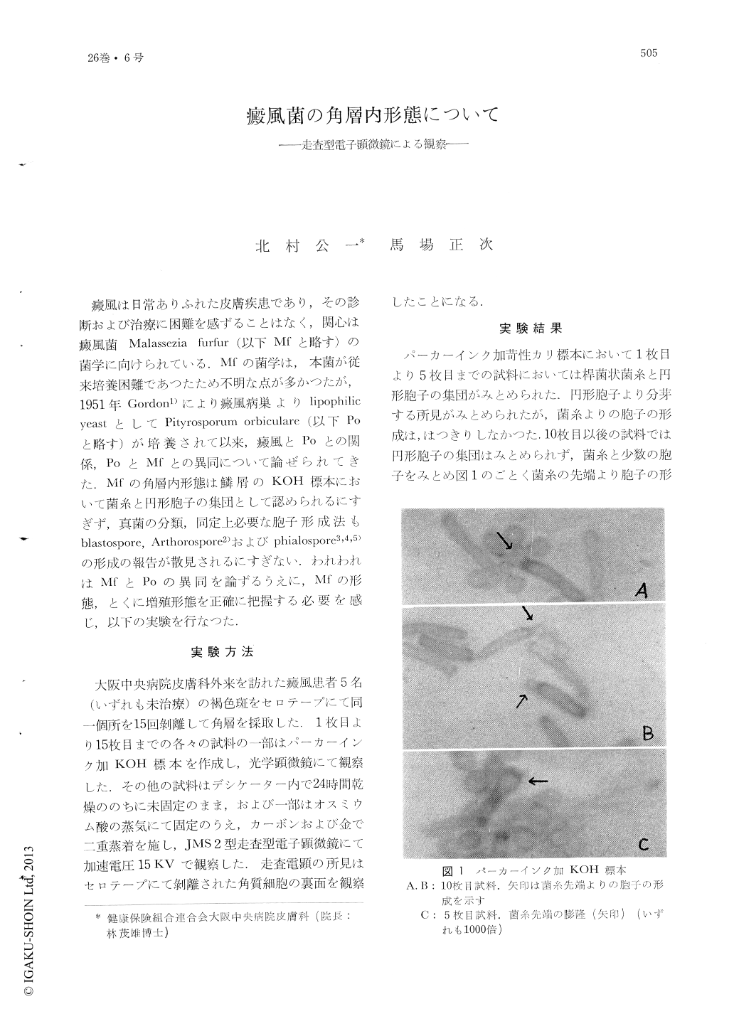
癜風は日常ありふれた皮膚疾患であり,その診断および治療に困難を感ずることはなく,関心は癜風菌Malassczia furfur (以下Mfと略す)の菌学に向けられている.Mfの菌学は,本菌が従来培養困難であつたため不明な点が多かつたが,1951年Gordon1)により癜風病巣よりlipophilic yeastとしてPityrosporum oibicularc (以下Poと略す)が培養されて以来,癜風とPoとの関係,PoとMfとの異同について論ぜられてきた,Mfの角層内形態は鱗屑のKOH標本において菌糸と円形胞子の集団として認められるにすぎず,真菌の分類,同定上必要な胞子形成法もblastospore, Arthorospore2)およびphialospore3,4,5)の形成の報告が散見されるにすぎない.われわれはMfとPoの異同を論ずるうえに,Mfの形態,とくに増殖形態を正確に把握する必要を感じ,以下の実験を行なつた.
Scales obtained with scotch tape from the lesion of tinea versicolor were observed by the scanning electron microscope. In the upper part of the horny layer the hyphac and the aggregated spherical spores were recognized. The spores showed the blastospore Formation by the budding, or the hyphae formation by germination. The latter showed branching and grew downward in the horny layer. The hyphae went through the horny cells, indicating keratolytic activity. The hyphac in the lower part of the horny layer formed the phialid and phialospore. The latter was carried upward in the process of the keratinization and again budding in the upper horny layer. It seemed that Malassezia furfur might show a morphological cycle in the horny layer. Formation of phialospores is important finding from the mycological standpoint.
Copyright © 1972, Igaku-Shoin Ltd. All rights reserved.