Japanese
English
- 有料閲覧
PPV購入案内
1,320円 (1,200円+税10%) こちらは、324ページから324ページ、339ページから344ページの文献です。購入には医書.jp本会員登録が必要です。
会員登録完了後に改めて上記「購入サイトへ」を選択してください。
または「購入サイトへ」選択後に「購入ログイン」、「新規会員登録」の順に進んでください。
会員登録について詳しくはをご確認ください。
- Abstract 文献概要
- 1ページ目 Look Inside
はじめに
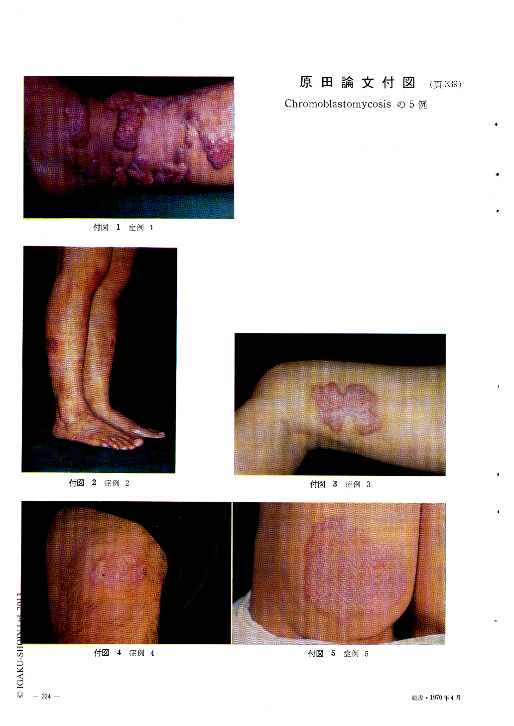
昭和36年より昭和43年までの間に当科において経験した黒色分芽菌症の5例について報告するとともに,いささか考察を加えたいと思う。
Five cases of this disease in the Department of Dermatology, Nippon Medical School, for 7 years-from 1961 to 1968-, were reported.
The results of the clinical, histological, and mycological studies are as follows :
1. The causative fungi were all Hormodendrum pedrosoi.
2. The clinical pictures of the lesions looked like tuberculosis of the skin, especially tuberculosis verrucosa cutis.
3. Macroscopic findings of one case were similar to that of impetigo, which seemed to be the early stage of this disease.
4. Their histologic pictures resembled also that of tuberculosis. From histologic as well as clinical findings this disease is liable to be erroneously diagnosed as skin tuberculosis.
5. Fungus element showed a spore-form in the deeper layer of the skin, while in the horny layer many hyphae-form elements were found by NaOH test of scales. This fact suggested that those might be due to the difference of the immunological state of the layer of the skin where the fungus invaded.
6. The best therapeutic method, so far, was the resection of the lesion.
Copyright © 1970, Igaku-Shoin Ltd. All rights reserved.