Japanese
English
- 有料閲覧
PPV購入案内
1,320円 (1,200円+税10%) こちらは、1433ページから1436ページの文献です。購入には医書.jp本会員登録が必要です。
会員登録完了後に改めて上記「購入サイトへ」を選択してください。
または「購入サイトへ」選択後に「購入ログイン」、「新規会員登録」の順に進んでください。
会員登録について詳しくはをご確認ください。
- Abstract 文献概要
- 1ページ目 Look Inside
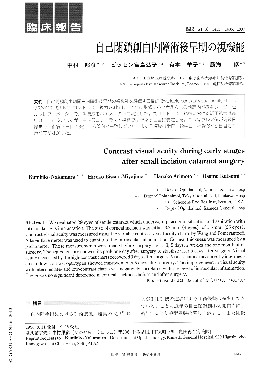
自己閉鎖創小切開白内障術後早期の視機能を評価する目的でvariable contrast visual acuity charts(VCVAC)を用いてコントラスト視力を測定し,これに影響すると考えられる前房内炎症をレーザーセルフレアーメーターで,角膜厚をパキメーターで測定した。高コントラスト視標における矯正視力は術後3日目に安定したが,中〜低コントラスト視標では術後5日目に安定した。これはフレア値が術翌日最高で,術後5日目で安定する傾向と一致していた。また角膜厚は術前,術翌日,術後3〜5日目で有意な差がなかった。
We evaluated 29 eyes of senile cataract which underwent phacoemulsification and aspiration with intraocular lens implantation. The size of corneal incision was either 3.2mm (4 eyes) of 5.5mm (25 eyes) . Contrast visual acuity was measured using the variable contrast visual acuity charts by Wang and Pomerantzeff. A laser flare meter was used to quantitate the intraocular inflammation. Corneal thickness was measured by a pachometer. These measurements were made before surgery and 1, 3, 5 days, 2 weeks and one month after surgery. The aqueous flare showed its peak one day after surgery to stabilize after 5 days after surgery. Visual acuity measured by the high-contrast charts recovered 3 days after surgery. Visual acuities measured by intermedi-ate- to low-contrast optotypes showed improvements 5 days after surgery. The improvement in visual acuity with intermediate- and low-contrast charts was negatively correlated with the level of intraocular inflammation. There was no significant difference in corneal thickness before and after surgery.
Copyright © 1997, Igaku-Shoin Ltd. All rights reserved.