Japanese
English
- 有料閲覧
PPV購入案内
1,320円 (1,200円+税10%) こちらは、625ページから628ページの文献です。購入には医書.jp本会員登録が必要です。
会員登録完了後に改めて上記「購入サイトへ」を選択してください。
または「購入サイトへ」選択後に「購入ログイン」、「新規会員登録」の順に進んでください。
会員登録について詳しくはをご確認ください。
- Abstract 文献概要
- 1ページ目 Look Inside
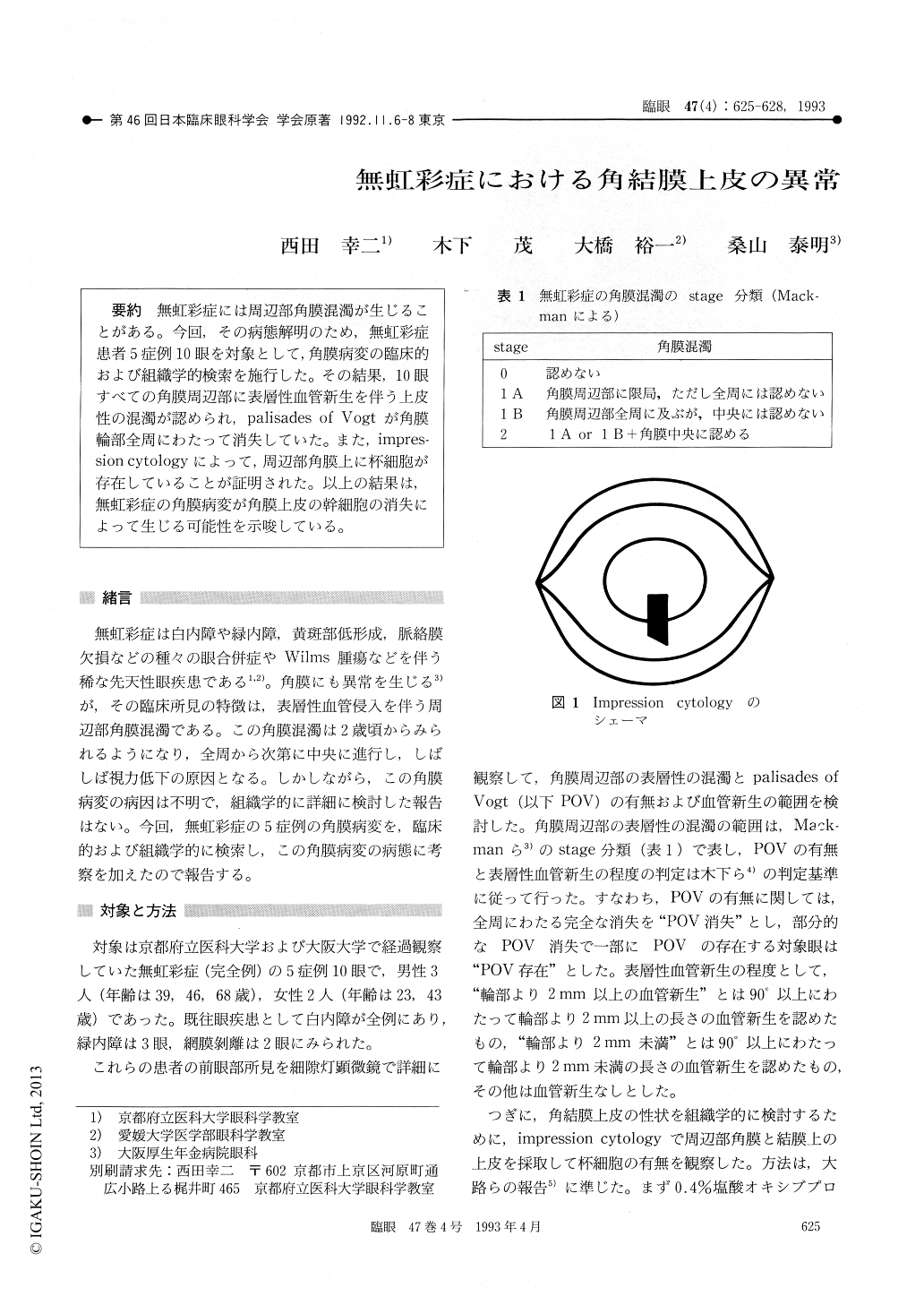
無虹彩症には周辺部角膜混濁が生じることがある。今回,その病態解明のため,無虹彩症患者5症例10眼を対象として,角膜病変の臨床的および組織学的検索を施行した。その結果,10眼すべての角膜周辺部に表層性血管新生を伴う上皮性の混濁が認められ,palisades of Vogtが角膜輪部全周にわたって消失していた。また,impres—sion cytologyによって、周辺部角膜上に杯細胞が存在していることが証明された。以上の結果は,無虹彩症の角膜病変が角膜上皮の幹細胞の消失によって生じる可能性を示唆している。
Aniridia is often associated, among others, with peripheral corneal opacity and vascularization of unknown etiology. We examined 5 patients with virtually total aniridia clinically and histologically with impression cytology. These patients com-prised 3 males aged 39, 46 and 68 years, and 2 females aged 23 and 43 years. Slitlamp examination showed epithelial and/or stromal opacification, and superficial vascularization in the peripheral cornea. Palisades of Vogt, which is normally located in the corneal limbus, were completely absent. Impression cytology showed numerous goblet cells in the cor-neal epithelium. These findings seemed to imply that corneal epithelial abnormalities in aniridia are secondary to depletion of stem cells in the corneal epithelium.
Copyright © 1993, Igaku-Shoin Ltd. All rights reserved.