Japanese
English
- 有料閲覧
PPV購入案内
1,320円 (1,200円+税10%) こちらは、505ページから510ページの文献です。購入には医書.jp本会員登録が必要です。
会員登録完了後に改めて上記「購入サイトへ」を選択してください。
または「購入サイトへ」選択後に「購入ログイン」、「新規会員登録」の順に進んでください。
会員登録について詳しくはをご確認ください。
- Abstract 文献概要
- 1ページ目 Look Inside
- 参考文献 Reference
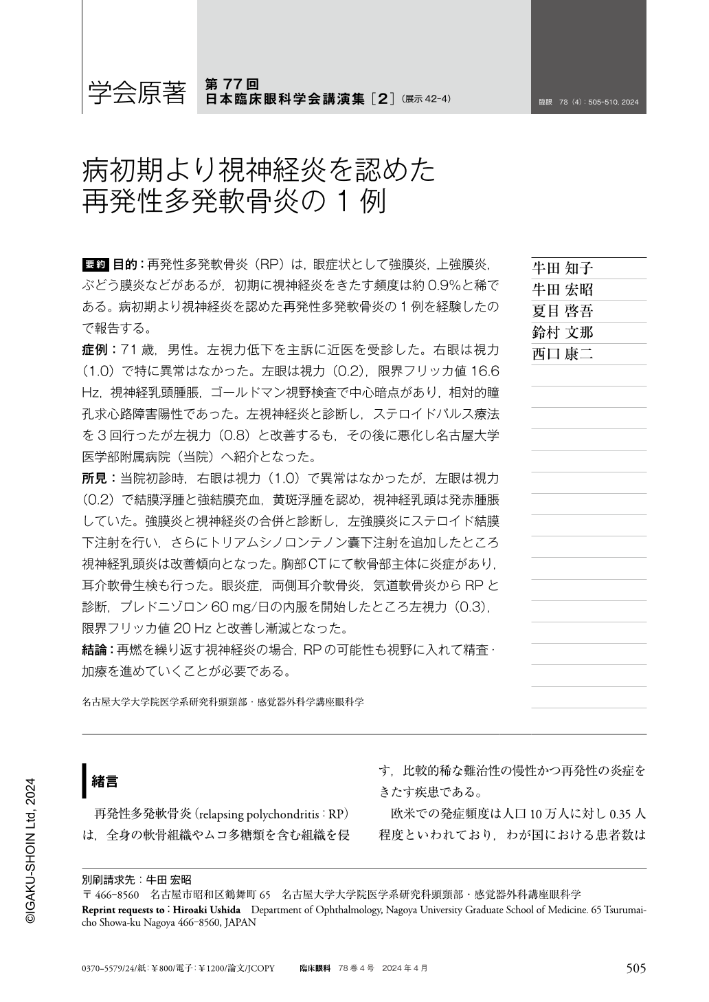
要約 目的:再発性多発軟骨炎(RP)は,眼症状として強膜炎,上強膜炎,ぶどう膜炎などがあるが,初期に視神経炎をきたす頻度は約0.9%と稀である。病初期より視神経炎を認めた再発性多発軟骨炎の1例を経験したので報告する。
症例:71歳,男性。左視力低下を主訴に近医を受診した。右眼は視力(1.0)で特に異常はなかった。左眼は視力(0.2),限界フリッカ値16.6Hz,視神経乳頭腫脹,ゴールドマン視野検査で中心暗点があり,相対的瞳孔求心路障害陽性であった。左視神経炎と診断し,ステロイドパルス療法を3回行ったが左視力(0.8)と改善するも,その後に悪化し名古屋大学医学部附属病院(当院)へ紹介となった。
所見:当院初診時,右眼は視力(1.0)で異常はなかったが,左眼は視力(0.2)で結膜浮腫と強結膜充血,黄斑浮腫を認め,視神経乳頭は発赤腫脹していた。強膜炎と視神経炎の合併と診断し,左強膜炎にステロイド結膜下注射を行い,さらにトリアムシノロンテノン囊下注射を追加したところ視神経乳頭炎は改善傾向となった。胸部CTにて軟骨部主体に炎症があり,耳介軟骨生検も行った。眼炎症,両側耳介軟骨炎,気道軟骨炎からRPと診断,プレドニゾロン60mg/日の内服を開始したところ左視力(0.3),限界フリッカ値20Hzと改善し漸減となった。
結論:再燃を繰り返す視神経炎の場合,RPの可能性も視野に入れて精査・加療を進めていくことが必要である。
Abstract Purpose:Ocular manifestations of recurrent polychondritis(RP)include scleritis, episcleritis, and uveitis, but optic neuritis is rare, occurring in only 0.9% of patients. We report a case of RP with optic neuritis as an early-onset manifestation.
Case:A 71-year-old man visited a clinic with a chief complaint of decreased visual acuity in his left eye. There were no abnormalities in his right eye. His left eye visual acuity was 0.2 with a left critical flicker fusion frequency(CFF)of 16.6 Hz and left optic disc swelling. His left eye had positive relative afferent pupillary defect, and Goldmann visual field testing showed a left central scotoma;accordingly, he was diagnosed with left optic neuritis and underwent steroid pulse therapy thrice. Although his left visual acuity improved to 0.8, it subsequently worsened and he was referred to our hospital.
Findings:At the time of the initial visit to our hospital, there was no visual loss in the right eye. However, the visual acuity of the left eye was 0.2. There was conjunctival edema, hyperemia of the sclera and conjunctiva, and macular edema in the left eye. In addition, the left optic nerve papilla was swollen. He was diagnosed with combined scleritis and optic neuritis. Subconjunctival steroid injections were administered for the left scleritis, and subtenon sac triamcinolone injections, were added. The optic neuritis in the left eye tended to improve. Chest CT showed inflammation mainly in the cartilage area, and an auricular cartilage biopsy was performed. He was diagnosed with RP owing to the presence of ocular inflammation, bilateral auricular chondritis, and airway chondritis. Oral prednisolone 60 mg was initiated, and left visual acuity improved to 0.3 and CFF improved to 20 Hz.
Conclusion:In cases of optic neuritis that repeatedly relapse, it is necessary to proceed with detailed examination and treatment, keeping in mind the possibility of RP.
Copyright © 2024, Igaku-Shoin Ltd. All rights reserved.