Japanese
English
- 有料閲覧
PPV購入案内
1,320円 (1,200円+税10%) こちらは、297ページから300ページの文献です。購入には医書.jp本会員登録が必要です。
会員登録完了後に改めて上記「購入サイトへ」を選択してください。
または「購入サイトへ」選択後に「購入ログイン」、「新規会員登録」の順に進んでください。
会員登録について詳しくはをご確認ください。
- Abstract 文献概要
- 1ページ目 Look Inside
- 参考文献 Reference
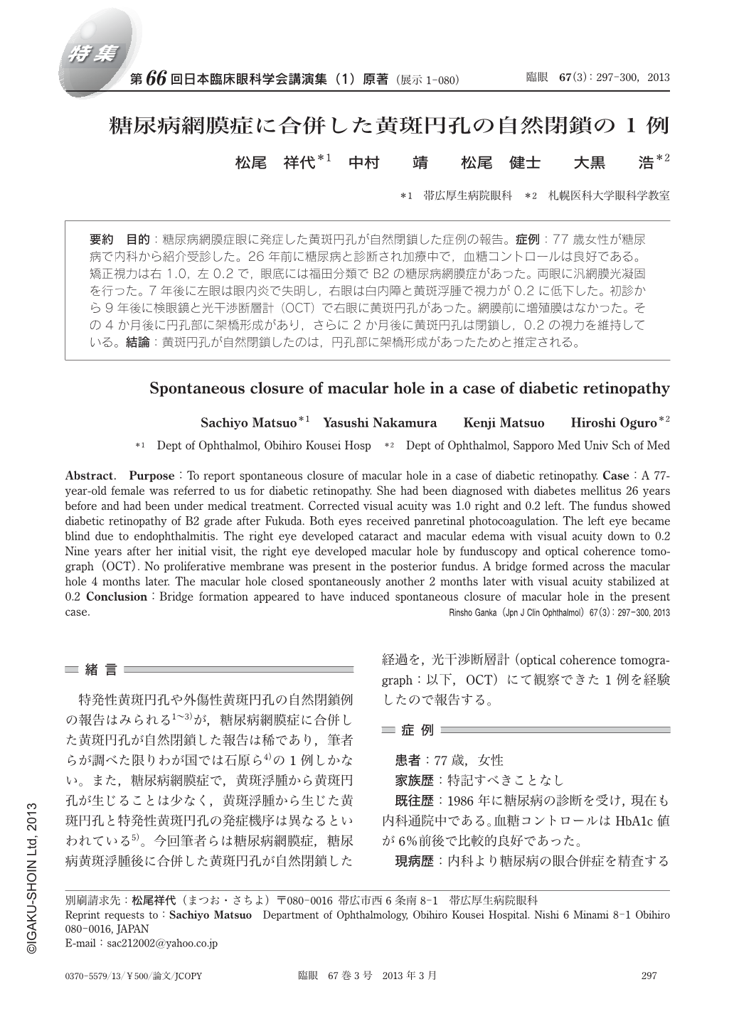
要約 目的:糖尿病網膜症眼に発症した黄斑円孔が自然閉鎖した症例の報告。症例:77歳女性が糖尿病で内科から紹介受診した。26年前に糖尿病と診断され加療中で,血糖コントロールは良好である。矯正視力は右1.0,左0.2で,眼底には福田分類でB2の糖尿病網膜症があった。両眼に汎網膜光凝固を行った。7年後に左眼は眼内炎で失明し,右眼は白内障と黄斑浮腫で視力が0.2に低下した。初診から9年後に検眼鏡と光干渉断層計(OCT)で右眼に黄斑円孔があった。網膜前に増殖膜はなかった。その4か月後に円孔部に架橋形成があり,さらに2か月後に黄斑円孔は閉鎖し,0.2の視力を維持している。結論:黄斑円孔が自然閉鎖したのは,円孔部に架橋形成があったためと推定される。
Abstract. Purpose:To report spontaneous closure of macular hole in a case of diabetic retinopathy. Case:A 77-year-old female was referred to us for diabetic retinopathy. She had been diagnosed with diabetes mellitus 26 years before and had been under medical treatment. Corrected visual acuity was 1.0 right and 0.2 left. The fundus showed diabetic retinopathy of B2 grade after Fukuda. Both eyes received panretinal photocoagulation. The left eye became blind due to endophthalmitis. The right eye developed cataract and macular edema with visual acuity down to 0.2 Nine years after her initial visit, the right eye developed macular hole by funduscopy and optical coherence tomograph(OCT). No proliferative membrane was present in the posterior fundus. A bridge formed across the macular hole 4 months later. The macular hole closed spontaneously another 2 months later with visual acuity stabilized at 0.2 Conclusion:Bridge formation appeared to have induced spontaneous closure of macular hole in the present case.
Copyright © 2013, Igaku-Shoin Ltd. All rights reserved.