Japanese
English
- 有料閲覧
PPV購入案内
1,320円 (1,200円+税10%) こちらは、1775ページから1779ページの文献です。購入には医書.jp本会員登録が必要です。
会員登録完了後に改めて上記「購入サイトへ」を選択してください。
または「購入サイトへ」選択後に「購入ログイン」、「新規会員登録」の順に進んでください。
会員登録について詳しくはをご確認ください。
- Abstract 文献概要
- 1ページ目 Look Inside
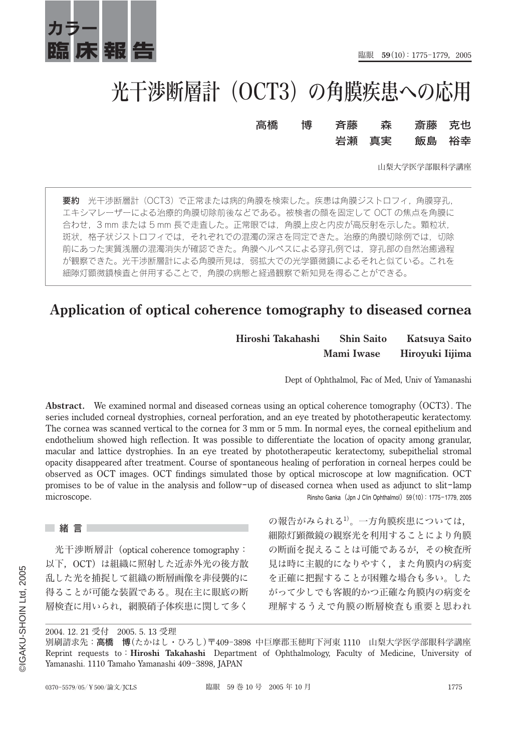
光干渉断層計(OCT3)で正常または病的角膜を検索した。疾患は角膜ジストロフィ,角膜穿孔,エキシマレーザーによる治療的角膜切除前後などである。被検者の顔を固定してOCTの焦点を角膜に合わせ,3mmまたは5mm長で走査した。正常眼では,角膜上皮と内皮が高反射を示した。顆粒状,斑状,格子状ジストロフィでは,それぞれでの混濁の深さを同定できた。治療的角膜切除例では,切除前にあった実質浅層の混濁消失が確認できた。角膜ヘルペスによる穿孔例では,穿孔部の自然治癒過程が観察できた。光干渉断層計による角膜所見は,弱拡大での光学顕微鏡によるそれと似ている。これを細隙灯顕微鏡検査と併用することで,角膜の病態と経過観察で新知見を得ることができる。
We examined normal and diseased corneas using an optical coherence tomography(OCT3). The series included corneal dystrophies,corneal perforation,and an eye treated by phototherapeutic keratectomy. The cornea was scanned vertical to the cornea for 3 mm or 5 mm. In normal eyes,the corneal epithelium and endothelium showed high reflection. It was possible to differentiate the location of opacity among granular,macular and lattice dystrophies. In an eye treated by phototherapeutic keratectomy,subepithelial stromal opacity disappeared after treatment. Course of spontaneous healing of perforation in corneal herpes could be observed as OCT images. OCT findings simulated those by optical microscope at low magnification. OCT promises to be of value in the analysis and follow-up of diseased cornea when used as adjunct to slit-lamp microscope.
Copyright © 2005, Igaku-Shoin Ltd. All rights reserved.