Japanese
English
- 有料閲覧
PPV購入案内
1,320円 (1,200円+税10%) こちらは、379ページから383ページの文献です。購入には医書.jp本会員登録が必要です。
会員登録完了後に改めて上記「購入サイトへ」を選択してください。
または「購入サイトへ」選択後に「購入ログイン」、「新規会員登録」の順に進んでください。
会員登録について詳しくはをご確認ください。
- Abstract 文献概要
- 1ページ目 Look Inside
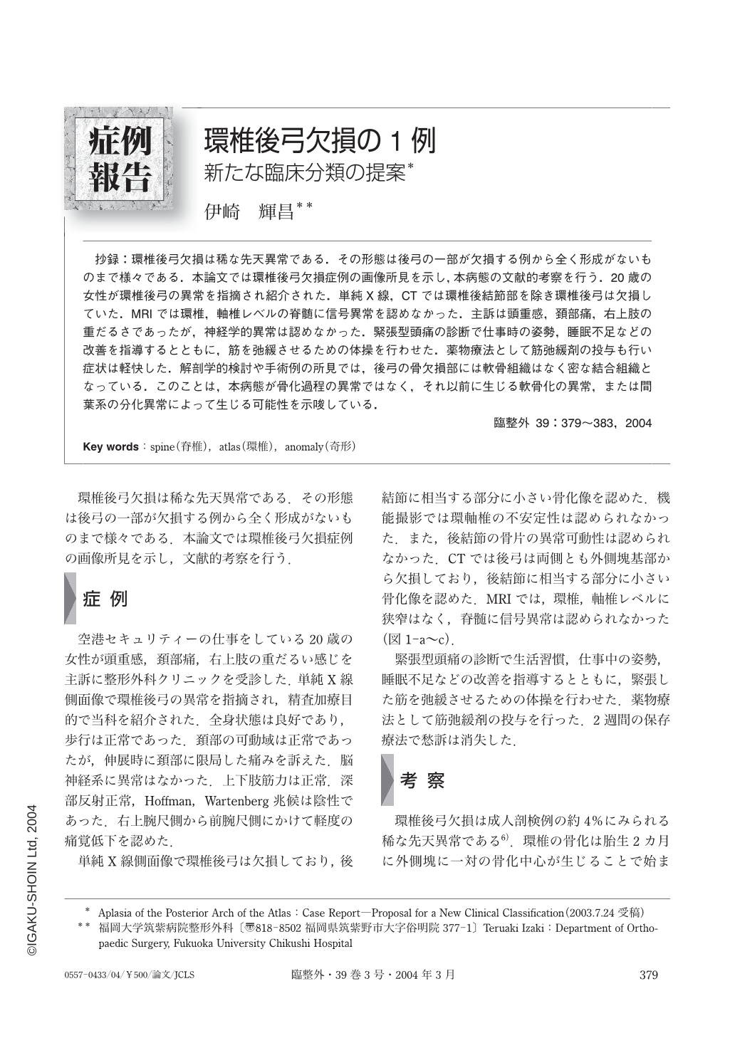
抄録:環椎後弓欠損は稀な先天異常である.その形態は後弓の一部が欠損する例から全く形成がないものまで様々である.本論文では環椎後弓欠損症例の画像所見を示し,本病態の文献的考察を行う.20歳の女性が環椎後弓の異常を指摘され紹介された.単純X線,CTでは環椎後結節部を除き環椎後弓は欠損していた.MRIでは環椎,軸椎レベルの脊髄に信号異常を認めなかった.主訴は頭重感,頚部痛,右上肢の重だるさであったが,神経学的異常は認めなかった.緊張型頭痛の診断で仕事時の姿勢,睡眠不足などの改善を指導するとともに,筋を弛緩させるための体操を行わせた.薬物療法として筋弛緩剤の投与も行い症状は軽快した.解剖学的検討や手術例の所見では,後弓の骨欠損部には軟骨組織はなく密な結合組織となっている.このことは,本病態が骨化過程の異常ではなく,それ以前に生じる軟骨化の異常,または間葉系の分化異常によって生じる可能性を示唆している.
Partial or complete aplasia of the posterior arch of the atlas is a rare anomaly. The purpose of this report is to describe the radiological findings in a case we encountered and to review the literature on congenital defects of the posterior arch of the atlas. A 20-year-old woman who had been found to have an abnormality of the posterior arch of the atlas was referred to our hospital for a further diagnostic work-up. She complained of nonspecific neck pain and headaches, but a neurological examination revealed normal findings. Roentgenograms and CT scans revealed absence of the posterior arch of the atlas except for the posterior tubercle. MR imaging showed no evidence of compression or signal changes in the cord at the C1-C2 level. The symptoms rapidly subsided in response to conservative treatment. Recent studies have shown that the bony gap in the posterior arch is bridged by loose connective tissue rather than cartilage, and thus the anomaly is thought to be attributable to defective cartilage development rather than a disorder of ossification per se.
Copyright © 2004, Igaku-Shoin Ltd. All rights reserved.