Japanese
English
- 有料閲覧
PPV購入案内
1,320円 (1,200円+税10%) こちらは、85ページから92ページの文献です。購入には医書.jp本会員登録が必要です。
会員登録完了後に改めて上記「購入サイトへ」を選択してください。
または「購入サイトへ」選択後に「購入ログイン」、「新規会員登録」の順に進んでください。
会員登録について詳しくはをご確認ください。
- Abstract 文献概要
- 1ページ目 Look Inside
症例呈示
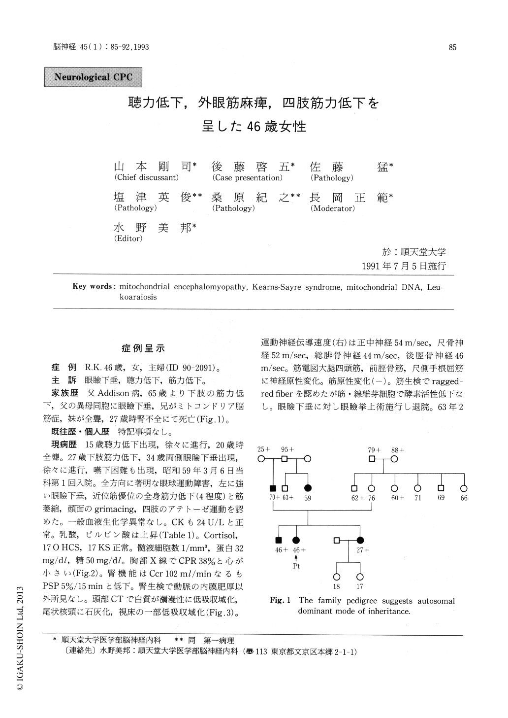
症例 R.K.46歳,女,主婦(ID 90-2091)。
主訴 眼瞼下垂,聴力低下,筋力低下。
We report a 46-year-old female who presented progressive ophthalmoplegia and limb weakness. She was well until the age of 15 years when there was an onset of bilateral deafness. She became completely deaf by 20 years of age. She noted an onset of weakness in her legs when she was 27-year -old, and of ptosis at 34 years of age. She was admitted to our hospital when she was 41-year-old. Neurological examination revealed near total ophthalmoplegia, bilateral ptosis, dysphagia, genera-lized muscle atrophy and weakness of approximate-ly 4/5 degree, facial grimacing, athetotic move-ments in four limbs. Laboratory examinations revealed increase in blood lactate and pyruvate levels and diffuse low density change in the cerebral white matter in CT scans. She was thought to have a mitochondrial encephalomyopathy. She was dis-charged for follow-up, but her clinical course wasthat of a relentless deterioration. She was readmit-ted to our service in December 1989. She showed further progress in her weakness and muscle atro-phy. Otherwise neurological examination was essen-tially similar to the previous one. Her cranial CT scans showed low density changes in striatum, thalamus and midbrain in addition to the white matter. Enzyme activities of the electron transport complexes revealed a moderate decrease in the succinatecytochrome c reductase activity, and the Southern blot analysis of mtDNA revealed multiple deletions in mitochondrial genomes. Two months after her admission, she developed bronchopneu-monia, and expired on March 13th, 1990.
Post-mortem examination revealed diffuse pallor of myeline in the cerebral white matter in K-B staining. A marked neuronal loss and gliosis were observed in putamen bilaterally. Skeletal muscles showed typical changes of mitochondrial myopath-ies with ragged-red fibers in Gomori-Trichrome staining, and crystalline inclusion bodies by electron microscopic observation. Some neurogenic atro-phies were also seen. Oculomotor nuclei appeared intact. It was thought that she had an incomplete form of Kearns-Sayre syndrome. The patient was discussed in a neurological CPC of the departments of Neurology and Pathology of Juntendo University School of Medicine.
Copyright © 1993, Igaku-Shoin Ltd. All rights reserved.