Japanese
English
- 有料閲覧
- Abstract 文献概要
- 1ページ目 Look Inside
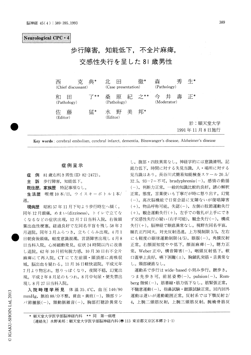
症例呈示
症例 81歳右利き男性(ID 82-2472)。
主訴 歩行障害,知能低下。
We present a 81-year old male who developed dementia, gait disturbance and right hemiparesis. He was well until the age of 74 when he developed a hemorrhagic infarction in the right occipital region, which left him left homonymous hemianop-sia. One year later he had one TIA attack consist-ing of dizziness, headache, and some clouding of consciousness. At that time, atrial fibrillation was found. At age 79, he was attacked by right hemipa-resis. Cranial CT scans revealed a lesion consistent with a hemorrhagic infarct in the left middle cere-bral artery territory. Two months prior to his final admission, he had a gradual onset of forgetfulness, labile affect, nocturnal agitation and hallucination which were followed by gait disturbance and uri-nary incontinence.
On admission, he was alert but moderately demented. In addition he showed difficulty in repeti-tion, limb kinetic and ideomotor apraxia of the left hand indicative of sympathetic apraxia, and con-structional apraxia bilaterally. Granial nerves appeared intact except for left homonymoushemianopsia. His gait was wide-based and small stepped. No weakness or ataxia was noted. Deep reflexes were diminished on the left side. Plantar reflex was equivocally extensor on the left. Light touch and pain was slightly diminished on the right side. Cranial CT scans revealed a large low density area in the left fronto-temporo-parietal region. Also ventricular dilatation, diffuse low densitychange in the subcortical white matter, and diffuse cortical atrophy were seen. His clinical course was complicated by melena, anemia, pneumonia, cardiac failure and renal failure. He expired 2 months after his admission.
Post-mortem examination revealed old cerebral infarctions one in the right posterior cerebral artery territory and one in the left middle cerebral artery distribution. Also pallor of myelin was noted in a myelin staining in both frontal and occipital areas bilaterally consistent with Binswanger's ence-phalopathy, however, arteriolosclerotic changes were not prominent in the white matter suggesting circulatory disturbance as an etiologic factor for the pallor of myelin. These old infarctions appeared in all likelihood of embolic nature because hemoside-rin was present in the infarcted areas suggesting hemorrhagic infarction. In addition, numerous senile plaques were seen all over the cerebral cor-tices. A small amount of neurofibrillary tangles were also seen in hippocampi. These findings were consistent with Alzheimer's disease. In summary this patient had multiple cerebral emboli, Binswanger's encephalopathy and senile dementia of Alzheimer type.

Copyright © 1993, Igaku-Shoin Ltd. All rights reserved.


