Japanese
English
- 有料閲覧
PPV購入案内
1,320円 (1,200円+税10%) こちらは、339ページから343ページの文献です。購入には医書.jp本会員登録が必要です。
会員登録完了後に改めて上記「購入サイトへ」を選択してください。
または「購入サイトへ」選択後に「購入ログイン」、「新規会員登録」の順に進んでください。
会員登録について詳しくはをご確認ください。
- Abstract 文献概要
- 1ページ目 Look Inside
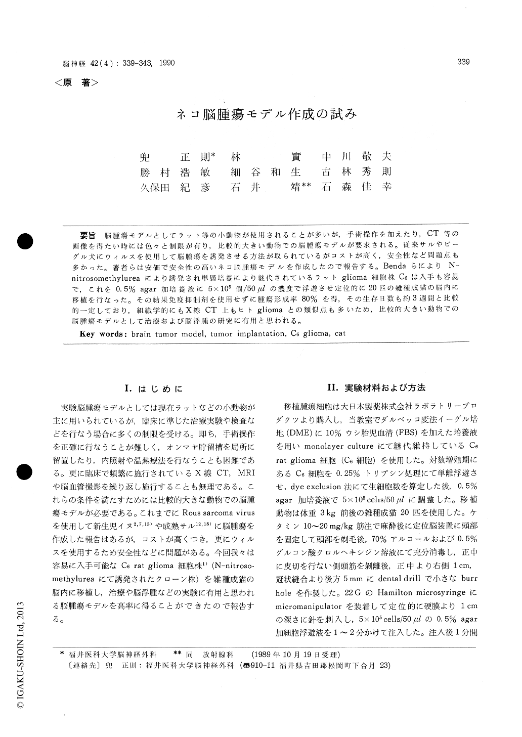
脳腫瘍モデルとしてラット等の小動物が使用されることが多いが,手術操作を加えたり,CT等の画像を得たい時には色々と制限が有り,比較的大きい動物での脳腫瘍モデルが要求される。従来サルやビーグル犬にウィルスを使用して脳腫瘍を誘発させる方法が取られているがコストが高く,安全性など問題点も多かった。著者らは安価で安全性の高いネコ脳腫瘍モデルを作成したので報告する。BendaらによりN—nitrosomethylureaにより誘発され単層培養により継代されているラットglioma細胞株C6は入手も容易で,これを0.5%agar加培養液に5×10⁵個/50μlの濃度で浮遊させ定位的に20匹の雑種成猫の脳内に移植を行なった。その結果免疫抑制剤を使用せずに腫瘍形成率80%を得,その生存日数も約3週間と比較的一定しており,組織学的にもX線CT上もヒトgliomaとの類似点も多いため,比較的人きい動物での脳腫瘍モデルとして治療および脳浮腫の研究に有用と思われる。
Small animal models such as the rat have seri-ous limitations for multiple human scale instru-mentations, surgical manipulations, and compute-rized tomographic (CT) evaluations, so that large animal models are required for the study using them. Although brain tumors induced with Rous sarcoma virus in neonatal beagle or adult monkey had been reported, these animals are very expen-sive ones for tumor research. A major drawback of virally induced brain tumor model is, moreo-ver, the need for specialized viral facilities and safety precautions for laboratory personnel. In this paper, a cat glioma model implanted with C6 glioma cells derived from rats injected with N-nit-rosomethylurea is reported. For an implantation dose of 5×10⁵ cells/50 μl. C6 glioma cells were suspended in modified Eagle medium supplemen-ted with 10% fetal bovine serum and 0.5% agar. Twenty adult mongrel cats were injected with 5 × 10⁵ C6 glioma cells intracerebrally. Implanted cats had brain tumors of about 10 mm in diameter with a yield of 80%. The mean survival was about 3 weeks after implantation. Tumors developed as spheroidal, hemorrhagic masses with central areas of necrosis and peripheral edema. They were lo-cated within the parenchyma of the implanted re-gion. This tumor possessed many of the histolo-gical and radiological characteristics of human glioblastoma such as the following : Areas of he-morrhage and necrosis surrounded by pseudopal-lisading were observed within the tumor consis-ting of spindle-shaped cells with pleomorphic nuc-lei. A mass lesion with ring or garland-like enhan-cement surrounded by brain edema was shown on the CT scans. This brain tumor models in cat has many advantages including low cost, reproducible morphology, a short survival time, safety for the investigator, and the feasibility for surgical mani-pulation and sequential CT scanning.
Copyright © 1990, Igaku-Shoin Ltd. All rights reserved.