Japanese
English
- 有料閲覧
- Abstract 文献概要
- 1ページ目 Look Inside
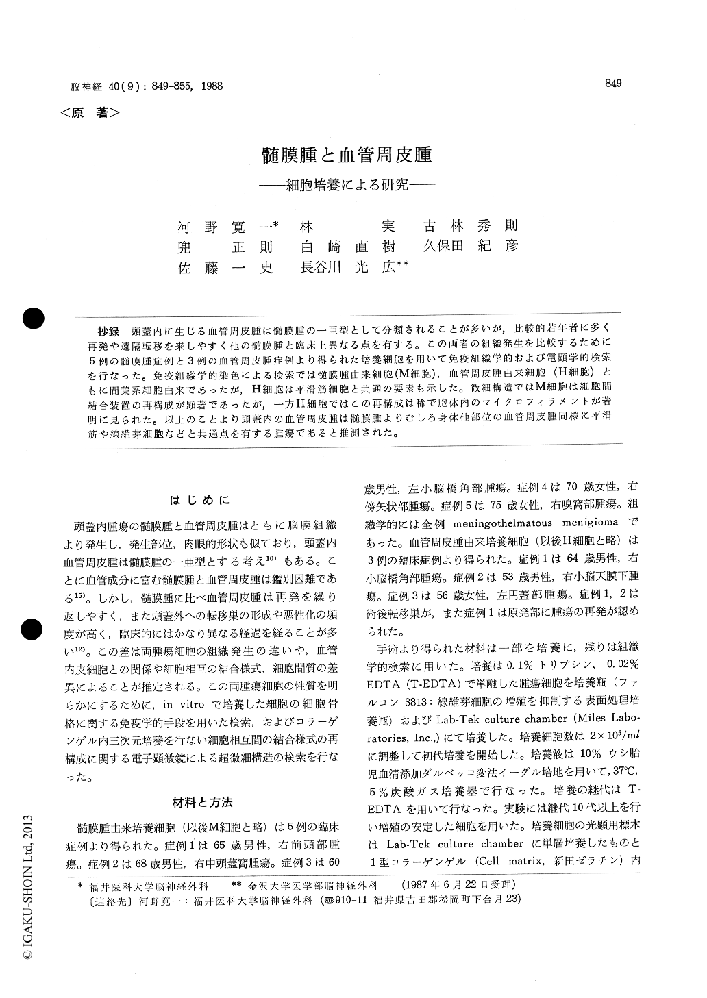
抄録 頭蓋内に生じる血管周皮腫は髄膜腫の一亜型として分類されることが多いが,比較的若年者に多く再発や遠隔転移を来しやすく他の髄膜腫と臨床上異なる点を有する。この両者の組織発生を比較するために5例の髄膜腫症例と3例の血管周皮腫症例より得られた培養細胞を用いて免疫組織学的および電顕学的検索を行なった。免疫組織学的染色による検索では髄膜腫由来細胞(M細胞),血管周皮腫由来細胞(H細胞)ともに間葉系細胞由来であったが,H細胞は平滑筋細胞と共通の要素も示した。微細構造ではM細胞は細胞間結合装置の再構成が顕著であったが,一方H細胞ではこの再構成は稀で胞体内のマイクロフィラメントが著明に見られた。以上のことより頭蓋内の血管周皮腫は髄膜腫よりむしろ身体他部位の血管周皮腫同様に平滑筋や線維芽細胞などと共通点を有する腫瘍であると推測された。
Intracranial meningioma cells and hemangioperi-cytoma cells were cultured in vitro for immuno-histological and ultrastructural study. Anti-vimen-tin, desmin, actin, a-actinin and laminin mono-clonal antibodies were applied for immunohisto-logical examination. Three dimension culture cells in collagen gel were examined by electron microscopy for ultrastructural studies.
Cultured meningioma cells and hemangioperi-cytoma cells were positive for anti-vimentin stain, while desmin, actin, a-actinin were posi-tive in only hemangiopericytoma cells. The culture morphology of these two cells was same in light microscopic study except for the whorl forma-tion in menigioma cells. Reconstruction of inter-cellular junctions such as desmosomes and junc-tional complexes were common in cultured menin-gioma cells but seldom in hemangiopericytoma cells. Hemangiopericytoma cells continued to have prominent intracytoplasmic intermediate filaments and microfilaments forming dense bodies in cul-ture.
These results suggest that intracranial hem-angiopericytoma has something in common with smooth muscle cell and fibroblast cells which forms the vascular and perivascular tissues, and it is distinguishable from meningioma.

Copyright © 1988, Igaku-Shoin Ltd. All rights reserved.


