Japanese
English
- 有料閲覧
- Abstract 文献概要
- 1ページ目 Look Inside
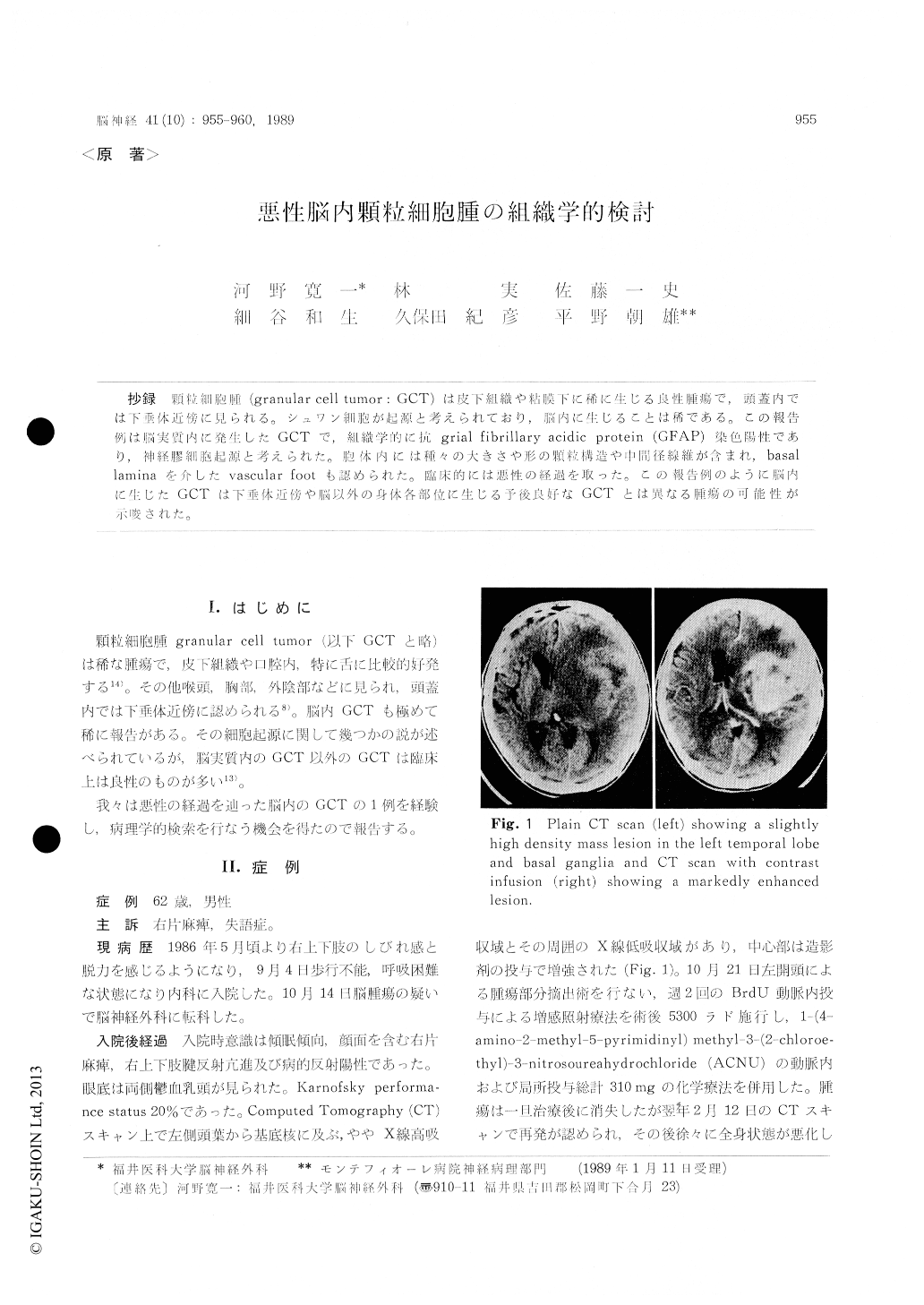
抄録 顆粒細胞腫(granular cell tumor: GCT)は皮下組織や粘膜下に稀に生じる良性腫瘍で,頭蓋内では下垂体近傍に見られる。シュワン細胞が起源と考えられており,脳内に生じることは稀である。この報告例は脳実質内に発生したGCTで,組織学的に抗grial fibrillary acidic protein (GFAP)染色陽性であり,神経膠細胞起源と考えられた。胞体内には種々の大きさや形の顆粒構造や中間径線維が含まれ,basallaminaを介したvascular footも認められた。臨床的には悪性の経過を取った。この報告例のように脳内に生じたGCTは下垂体近傍や脳以外の身体各部位に生じる予後良好なGCTとは異なる腫瘍の可能性が示唆された。
Granular cell tumor (GCT), which is suspected to be of Schwann cell origin, sometimes grows in the subcutaneous tissue, oral cavity and visceral sites and this tumor has a rather benign nature. Intracranial GCT also grows in the neurohypo-physis but rarely in the brain parenchyma. We reported a case of intra-cerebral GCT in the left hemisphere, which took a malignant course. The patient was a 62-year-old male with a history of slowly progressing right hemiparesis and aphasia since May 1986. He was in a drowsy state and showed right hemiplegia on admission (October 14, 1986). Radiological examinations revealed a tumor and surrounding edema in the left temporal lobe and basal ganglias. Resection of the tumor and both radiotherapy of 53 Grey and chemothe-rapy using ACNU (total 310 mg) and BrdU (500 mg, two times per week prior to radiation) were applied after the operation. Although the tumordisappeared once after these treatments, the pati-ent died of recurrence on July 3, 1987. Histolo-gical examinations on the specimen taken at the first operation revealed that the tumor consisted of rather round, large and small cells with a few cell processes. The large cells often had bizarre and multiple nuclei. These large cells had rich eosinophilic granular particles of various size and vacuoles in their cytoplasm. The staining for anti-glial fibrillary acidic protein (GFAP) was positive in a part of the cytoplasm and cell processes. Elect-ron microscopically various sized and shaped gra-nular structures and intermediate filaments were noticed in the cytoplasm of both large and smal-ler cells. These granular structures, which had unit membrane, were observed to have changed to va-cuoles in other cells. It was evident that the vas-cular foot of the granular cell was in contact with pericytes by basal lamina. These findings indicated that this granular tumor is of astrocyte origin. The present report suggests that the intracerebral GCT such as in our case possibly belongs to a different tumor group from those which are found in the subcutaneous, submucosal tissues and the neurohypophysis which are suspected to be of Schwann cell origin
Copyright © 1989, Igaku-Shoin Ltd. All rights reserved.