Japanese
English
- 有料閲覧
PPV購入案内
1,320円 (1,200円+税10%) こちらは、199ページから203ページの文献です。購入には医書.jp本会員登録が必要です。
会員登録完了後に改めて上記「購入サイトへ」を選択してください。
または「購入サイトへ」選択後に「購入ログイン」、「新規会員登録」の順に進んでください。
会員登録について詳しくはをご確認ください。
- Abstract 文献概要
- 1ページ目 Look Inside
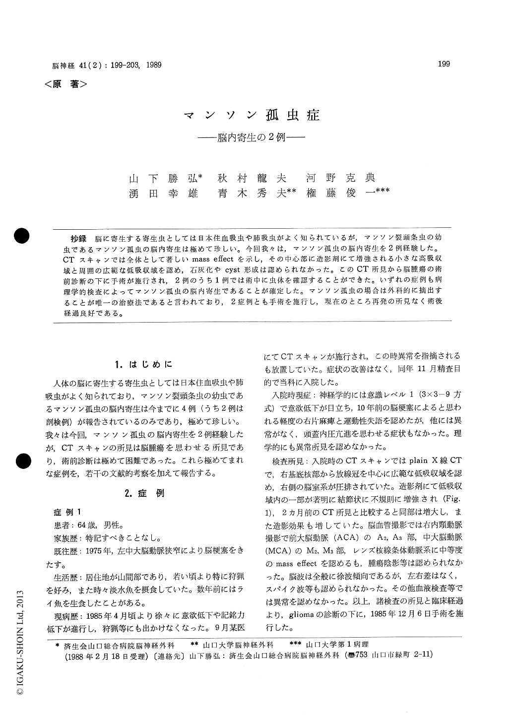
抄録 脳に寄生する寄生虫としては日本住血吸虫や肺吸虫がよく知られているが,マンソン裂頭条虫の幼虫であるマンソン孤虫の脳内寄生は極めて珍しい。今回我々は,マンソン孤虫の脳内寄生を2例経験した。CTスキャンでは全体として著しいmass effectを示し,その中心部に造影剤にて増強される小さな高吸収域と周囲の広範な低吸収域を認め,石灰化やcyst形成は認められなかった。このCT所見から脳腫瘍の術前診断の下に手術が施行され,2例のうち1例では術中に虫体を確認することができた。いずれの症例も病理学的検査によってマンソン孤虫の脳内寄生であることが確定した。マンソン孤虫の場合は外科的に摘出することが唯一の治療法であると言われており,2症例とも手術を施行し,現在のところ再発の所見なく術後経過良好である。
Cerebral Sparganosis Mansoni is a very rare disease caused by a larva of Diphyllobothrium and so far, only two cases have been reported. Another two cases are presented in this paper.
Case 1: A 64-year-old man was admitted pre-senting with hypovolition. He has eaten thunder fishes several years before. CT scan revealed an extensive low density area with an enhanced nod-ular mass around the right basal ganglia. An operation was carried out on the preoperative diagnosis of brain tumor and a granuloma was completely removed. His postoperative course was excellent. The histological examination confirmed the diagnosis of cerebral sparganosis.
Case 2: The patients was 60-year-old male showing hypesthesia in the left half of his face. He has often been to South Korea where patients of Sparganosis Mansoni are often reported. En-hanced CT scan demonstrated a small nodular region in the tip of the left temporal lobe. Spar-ganum Mansoni was identified during surgery and a granuloma was also extirpated. His postope-rative course was also excellent.
In cases with cerebral parasitism by Sparganum Mansoni, CT scan shows an extensive low density area with a nodular enhancement, but no calcifica-tion nor cyst formation. It is quite difficult to differenciate sparganosis from cerebral tumors on CT and labolatory data preoperatively. The dia-gnosis is usually confirmed by postoperative his-tological examination. As for the treatment of this disease, no effective chemotherapy is now available and surgical excision is recommended. Actually, four cases with cerebral sparganosis including two of our own demonstrated a satisfac-tory prognosis by surgical treatments.
Copyright © 1989, Igaku-Shoin Ltd. All rights reserved.