Japanese
English
- 有料閲覧
PPV購入案内
1,320円 (1,200円+税10%) こちらは、475ページから480ページの文献です。購入には医書.jp本会員登録が必要です。
会員登録完了後に改めて上記「購入サイトへ」を選択してください。
または「購入サイトへ」選択後に「購入ログイン」、「新規会員登録」の順に進んでください。
会員登録について詳しくはをご確認ください。
- Abstract 文献概要
- 1ページ目 Look Inside
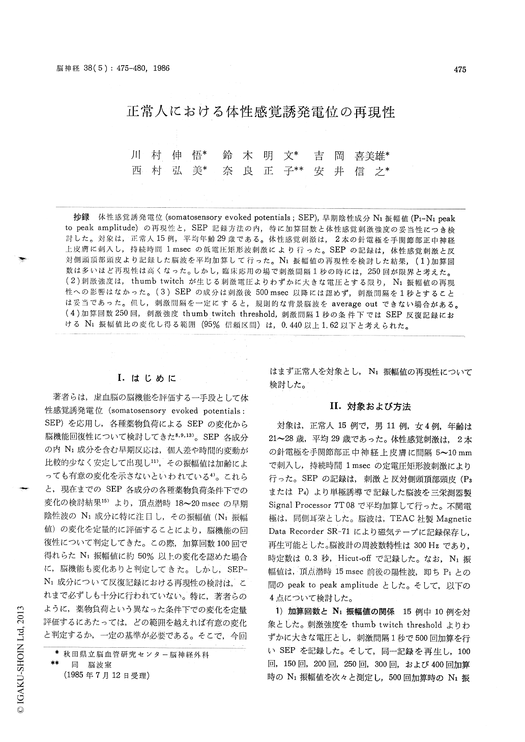
抄録 体性感覚誘発電位(somatosensory evoked potentials:SEP),早期陰性成分N1振幅値(P1-N1 peakto peak amplitude)の再現性と,SEP記録方法の内,特に加算回数と体性感覚刺激強度の妥当性につき検討した。対象は,正常人15例,平均年齢29歳である。体性感覚刺激は,2本の針電極を手関節部正中神経上皮膚に刺入し,持続時閥1msecの低電圧矩形波刺激により行った。SEPの記録は,体性感覚刺激と反対側頭頂部頭皮より記録した脳波を平均加算して行った。N1振幅値の再現性を検討した結果,(1)加算回数は多いほど再現性は高くなった。しかし,臨床応用の場で刺激間隔1秒の時には,250回が限界と考えた。(2)刺激強度は,thumb twitchが生じる刺激電圧よりわずかに大きな電圧とする限り,N1振幅値の再現性への影響はなかった。(3) SEPの成分は刺激後500msec以降には認めず,刺激間隔を1秒とすることは妥当であった。但し,刺激間隔を一定にすると,規則的な背景脳波をaverage outできない場合がある。(4)加算回数250回,刺激強度thumb twitch threshold,刺激間隔1秒の条件下ではSEP反復記録におけるN1振幅値比の変化し得る範囲(95%信頼区間)は,0.440以上1.62以下と考えられた。
The authors have applied the somatosensory evoked potentials (SEP) as an index of the brain function, and studied the reversibillity of the ischemic brain dysfunction from the SEP changes under the drug-induced conditions. From the pre-vious experiences, the authors are now doing the quantitative analysis of the amplitude of the early negative component (N1-amplitude) on SEP, and evaluating the reversibility of the brain dys-function. The early components of SEP, including N1 were reported that responses were reasonably constant in normal subjects. Responses recorded at different times in the same subject were usually very similar and there were not significant dif-ferences among the responses recorded in different subjects. In the consecutive SEP recordings, how-ever, the reproducibility of N1-amplitude has not yet fully studied. It is necessary to know the standard values when the significant changes of N1-amplitude are evaluated under the drug-induced conditions.
The purpose of this paper is to study the repro-ducibility of N1-amplitude of SEP in the consecu-tive recordings, and also, stimulating and record-ing methods of SEP. Subjects were 15 normal volunteers, from 21 to 38 (mean : 29) years old. Two stimulating needle electrodes were inserted to the skin at the wrist, and the somatosensory stimulation was applied to the median nerve with 1 msec duration of square pulse. Monopolar EEGs were recorded from the scalp of the parietal re-gion (P3 or P4) contralateral to the stimulation, and SEPs were recorded by averaging those EEGs with computer technique. N1-amplitude was deter-mined by the vertical distance between peaks of P1 and N1 components.
The results were as follows : 1) The reproducibility of N1-amplitude in the consecutive recordings was dependent on the fre-quency of averaging summation. But, it is difficult to record constant EEG for a long period because of motion artifacts or sleep. And the authors considered the limit 5 minutes. So, if stimulation pulses were generated every one second regularly, the limit of the frequency was considered 250-300 times.
2) The stimulus intensity did not have an influ-ence on the reproducibility of N1-amplitude, if the stimulus intensity was adjusted nearly above the thumb twitch threshold.
3) Components of SEP could not be recognized 500 msec after the somatosensory stimulation. Therefore, the stimulus interval of 1 sec was reasonable. With the regular stimulation, however, rhythmic EEG waves could not be averaged out.
4) In the consecutive SEP recordings by ave-raging 250 times every one second with the stimulus intensity of the thumb twitch threshold, N1-amplitude ratio, which compared Ni-amplitude of the first record with that of the second or third record, revealed between 0.440 and 1.62 (p <O.05).
Copyright © 1986, Igaku-Shoin Ltd. All rights reserved.