Japanese
English
- 有料閲覧
PPV購入案内
1,320円 (1,200円+税10%) こちらは、1101ページから1107ページの文献です。購入には医書.jp本会員登録が必要です。
会員登録完了後に改めて上記「購入サイトへ」を選択してください。
または「購入サイトへ」選択後に「購入ログイン」、「新規会員登録」の順に進んでください。
会員登録について詳しくはをご確認ください。
- Abstract 文献概要
- 1ページ目 Look Inside
I.はじめに
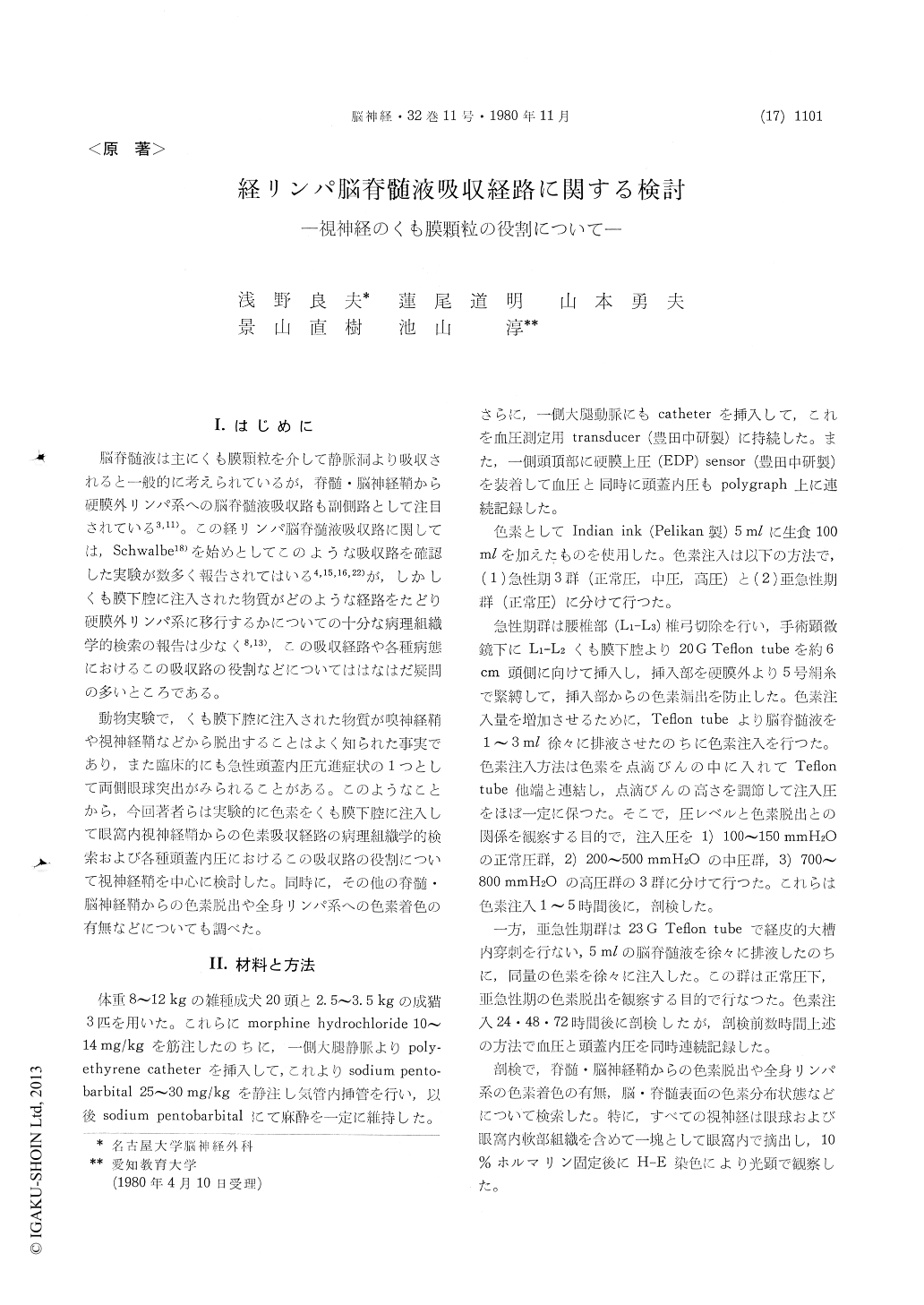
脳脊髄液は主にくも膜顆粒を介して静脈洞より吸収されると一般的に考えられているが,脊髄・脳神経鞘から硬膜外リンパ系への脳脊髄液吸収路も副側路として注目されている3,11)。この経リンパ脳脊髄液吸収路に関しては,Schwalbe18)を始めとしてこのような吸収路を確認した実験が数多く報告されてはいる4,15,16,22)が,しかしくも膜下腔に注入された物質がどのような経路をたどり硬膜外リンパ系に移行するかについての十分な病理組織学的検索の報告は少なく8,13),この吸収経路や各種病態におけるこの吸収路の役割などについてははなはだ疑問の多いところである。
動物実験で,くも膜下腔に注入された物質が嗅神経鞘や視神経鞘などから脱出することはよく知られた事実であり,また臨床的にも急性頭蓋内圧亢進症状の1つとして両側眼球突出がみられることがある。このようなことから,今回著者らは実験的に色素をくも膜下腔に注入して眼窩内視神経鞘からの色素吸収経路の病理組織学的検索および各種頭蓋内圧におけるこの吸収路の役割について視神経鞘を中心に検討した。同時に,その他の脊髄・脳神経鞘からの色素脱出や全身リンパ系への色素着色の有無などについても調べた。
It has been generally accepted that one of the CSF absorptive routes is from the subarachnoid space of the spinal and cranial nerves into the lymphatic system, but the precise pathway is not well understood.
Indian ink was introduced into the spinal sub-arachnoid space in 15 dogs and 3 cats to study the absorptive pathway of dye through the optic nerve sheath into the orbital tissue. These animals were killed within 5 hours after injecting the dye. Dur-ing each infusion period, ICP was monitored con-tinuously and was controlled at normal (100-150mmH2O), medium (200-500mmH2O) and high (700-800mmH2 O) pressure, respectively.
Table 1 shows the macroscopical findings in this study. At the high CSF pressure, the dye passed into the orbital tissue and the mucosa, mandibular and medial retropharyngeal lymph nodes were also stained in all specimens examined, but a few at the medium pressure. At the normal pressure in acute stage, the dye was not recognized on basal surface of the brain.
In 5 dogs, therefore, Indian ink of 5m1 was in-troduced after withdrawing an equal volume of CSF through the cisterna magna. These animals were killed in 24-72 hours of subacute stage, and ICP was normal.
The arachnoid villi were proved to be present in all specimens of optic nerves in the orbital portion by the microscopical examination. These arachnoid villi were classified into the three types, that is, projecting into the subdural space, intra-dural space and orbital tissue. At the high and medium CSF pressure, the dye passed from the subarachnoid space of the optic nerve through the arachnoid villi into the orbital tissue. At the normal CSF pressure in acute stage, the dye was not recognized into the optic nerve sheath. But in subacute stage, the dye was observed into thesubdural and intradural space of the optic nerve in the orbital portion and even into the orbital tissue microscopically.
Our observations indicate that the dye introduced into the subarachnoid space passed through the arachnoid villi not only under condition of increased ICP, but also under condition of normal ICP in 24-72 hours after injecting the dye. And under condition of increased ICP, the diffusion of dye was directly related to the pressure-dependent. These results suggested that the arachnoid villi acts a safety valve in regulating the CSF pressure, and the dye, which passed from the subarachnoid space of optic nerve through the arrchnoid villi into the orbital tissue, is absorpted into the lymphatic sys-tem.
Copyright © 1980, Igaku-Shoin Ltd. All rights reserved.