Japanese
English
- 有料閲覧
PPV購入案内
1,320円 (1,200円+税10%) こちらは、1266ページから1272ページの文献です。購入には医書.jp本会員登録が必要です。
会員登録完了後に改めて上記「購入サイトへ」を選択してください。
または「購入サイトへ」選択後に「購入ログイン」、「新規会員登録」の順に進んでください。
会員登録について詳しくはをご確認ください。
- Abstract 文献概要
- 1ページ目 Look Inside
- 参考文献 Reference
- サイト内被引用 Cited by
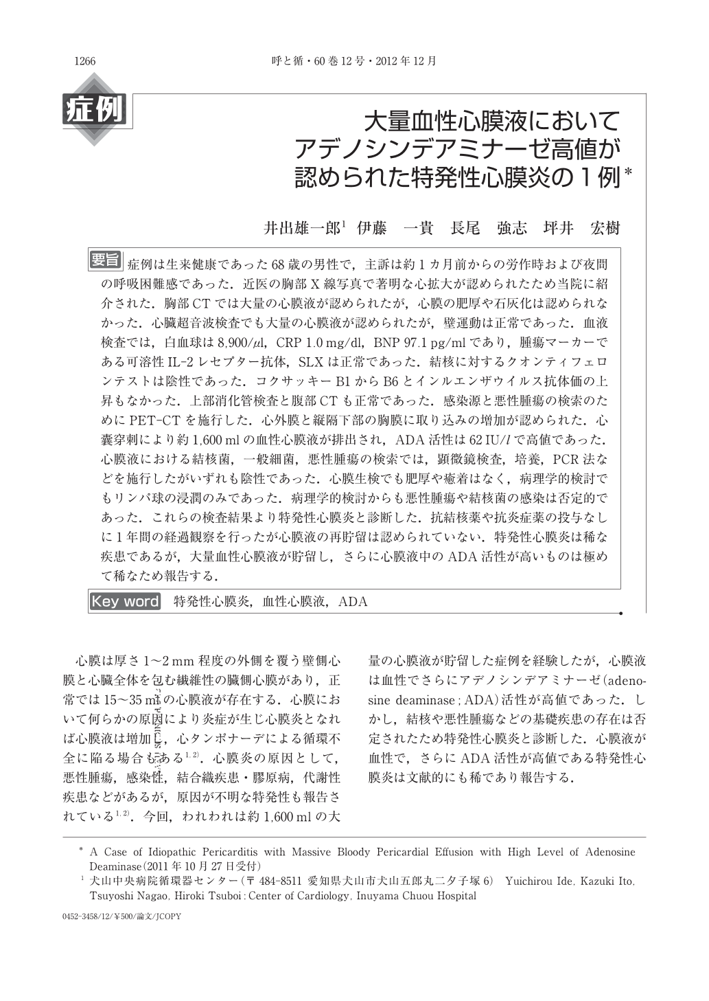
要旨 症例は生来健康であった68歳の男性で,主訴は約1カ月前からの労作時および夜間の呼吸困難感であった.近医の胸部X線写真で著明な心拡大が認められたため当院に紹介された.胸部CTでは大量の心膜液が認められたが,心膜の肥厚や石灰化は認められなかった.心臓超音波検査でも大量の心膜液が認められたが,壁運動は正常であった.血液検査では,白血球は8,900/μl,CRP 1.0mg/dl,BNP 97.1pg/mlであり,腫瘍マーカーである可溶性IL-2レセプター抗体,SLXは正常であった.結核に対するクオンティフェロンテストは陰性であった.コクサッキーB1からB6とインルエンザウイルス抗体価の上昇もなかった.上部消化管検査と腹部CTも正常であった.感染源と悪性腫瘍の検索のためにPET-CTを施行した.心外膜と縦隔下部の胸膜に取り込みの増加が認められた.心囊穿刺により約1,600mlの血性心膜液が排出され,ADA活性は62IU/lで高値であった.心膜液における結核菌,一般細菌,悪性腫瘍の検索では,顕微鏡検査,培養,PCR法などを施行したがいずれも陰性であった.心膜生検でも肥厚や癒着はなく,病理学的検討でもリンパ球の浸潤のみであった.病理学的検討からも悪性腫瘍や結核菌の感染は否定的であった.これらの検査結果より特発性心膜炎と診断した.抗結核薬や抗炎症薬の投与なしに1年間の経過観察を行ったが心膜液の再貯留は認められていない.特発性心膜炎は稀な疾患であるが,大量血性心膜液が貯留し,さらに心膜液中のADA活性が高いものは極めて稀なため報告する.
The patient was a 68-year-old man, who consulted our hospital complaining of dyspnea on effort and at night. Chest X-ray demonstrated marked cardiomegaly and chest computed tomography showed large amounts of pericardial effusion without pericardial thickening or calcification. Echocardiogram showed normal left ventricular wall motion without hemodynamic compromise. Blood examination confirmed a WBC count of 8,900/μl, CRP of 1.0mg/ml and a BNP of 97.1pg/ml. The tumor markers soluble IL-2 receptor and SLX were normal. A QuantiFERON test for Mycobacterium tuberculosis was negative. Viral titers of Coxsackie B1~B6 and influenza were not elevated during hospitalization. Upper gastrointestinal fiberscope examination and abdominal computed tomography were normal. To investigate septic focus or possible malignancy, 18F-FDG-PET-CT examination was performed. PET-CT examination showed high 18F-FDG uptake in the pericardium and in the pleura of the inferior mediastinum. Drainage of pericardial effusion was performed, and a total of 1,600ml of bloody pericardial effusion was excreted. The adenosine deaminase(ADA)value of pericardial effusion was 62IU/l, but smear and culture tests for M. tuberculosis, bacteria and malignancy were all negative.
The pericardium was resected, and biopsy showed only infiltration of lymphocytes with no adhesion or pericardial thickening. These pathological findings suggested no malignancy or infection by M. tuberculosis. These results suggest that the massive bloody pericardial effusion in this patient was the result of idiopathic pericarditis. Therefore, we observed this patient without antibiotics or anti-tuberculous therapy. Serial chest computed tomography showed no evidence of a relapse over a 12-month period after onset.
Copyright © 2012, Igaku-Shoin Ltd. All rights reserved.