Japanese
English
- 有料閲覧
PPV購入案内
1,320円 (1,200円+税10%) こちらは、529ページから532ページの文献です。購入には医書.jp本会員登録が必要です。
会員登録完了後に改めて上記「購入サイトへ」を選択してください。
または「購入サイトへ」選択後に「購入ログイン」、「新規会員登録」の順に進んでください。
会員登録について詳しくはをご確認ください。
- Abstract 文献概要
- 1ページ目 Look Inside
- 参考文献 Reference
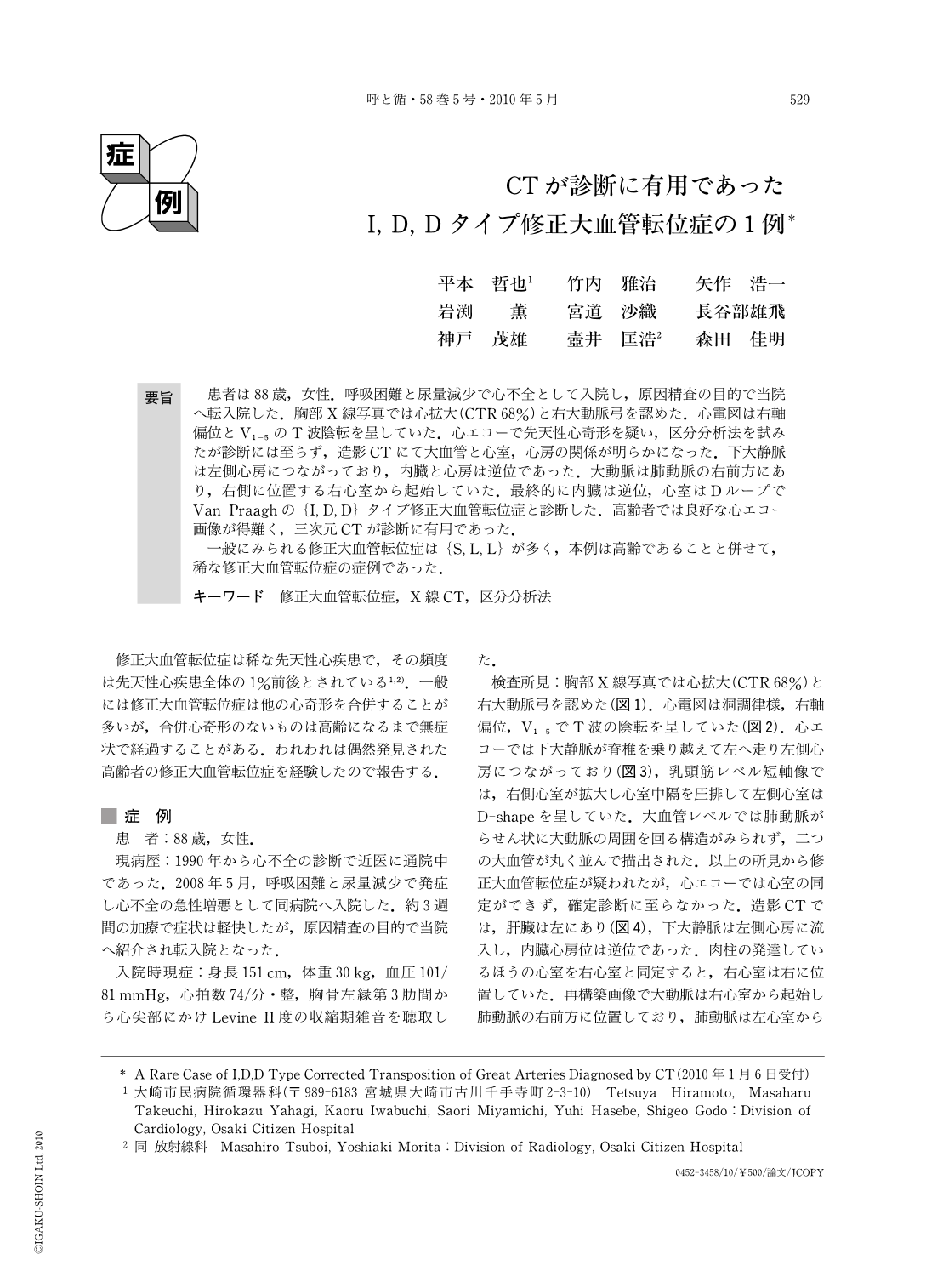
要旨 患者は88歳,女性.呼吸困難と尿量減少で心不全として入院し,原因精査の目的で当院へ転入院した.胸部X線写真では心拡大(CTR68%)と右大動脈弓を認めた.心電図は右軸偏位とV1-5のT波陰転を呈していた.心エコーで先天性心奇形を疑い,区分分析法を試みたが診断には至らず,造影CTにて大血管と心室,心房の関係が明らかになった.下大静脈は左側心房につながっており,内臓と心房は逆位であった.大動脈は肺動脈の右前方にあり,右側に位置する右心室から起始していた.最終的に内臓は逆位,心室はDループでVan Praaghの{I, D, D}タイプ修正大血管転位症と診断した.高齢者では良好な心エコー画像が得難く,三次元CTが診断に有用であった.
一般にみられる修正大血管転位症は{S, L, L}が多く,本例は高齢であることと併せて,稀な修正大血管転位症の症例であった.
An 88-year-old female was admitted complaining of dyspnea and oliguria. ECG showed right axis deviation and T wave inversion in V1-5. Chest X-ray revealed cardiomegaly and right side aortic arch. Congenital heart disease was suspected but segmental approach using echocardiography was not able to diagnose her clearly. Relationships among atria, ventricles and great arteries were detected clearly by X-ray CT. Finally, her condition was diagnosed as {I,D,D} type corrected transposition of great arteries. In older patient echocardiography often has problems in obtaining good images but CT, especially 3-D reconstruction, is useful for diagnosing congenital heart disease. This case is thought to be a rare case because of her high age and its anatomical classification.
Copyright © 2010, Igaku-Shoin Ltd. All rights reserved.