Japanese
English
- 有料閲覧
PPV購入案内
1,320円 (1,200円+税10%) こちらは、1139ページから1151ページの文献です。購入には医書.jp本会員登録が必要です。
会員登録完了後に改めて上記「購入サイトへ」を選択してください。
または「購入サイトへ」選択後に「購入ログイン」、「新規会員登録」の順に進んでください。
会員登録について詳しくはをご確認ください。
- Abstract 文献概要
- 1ページ目 Look Inside
- 参考文献 Reference
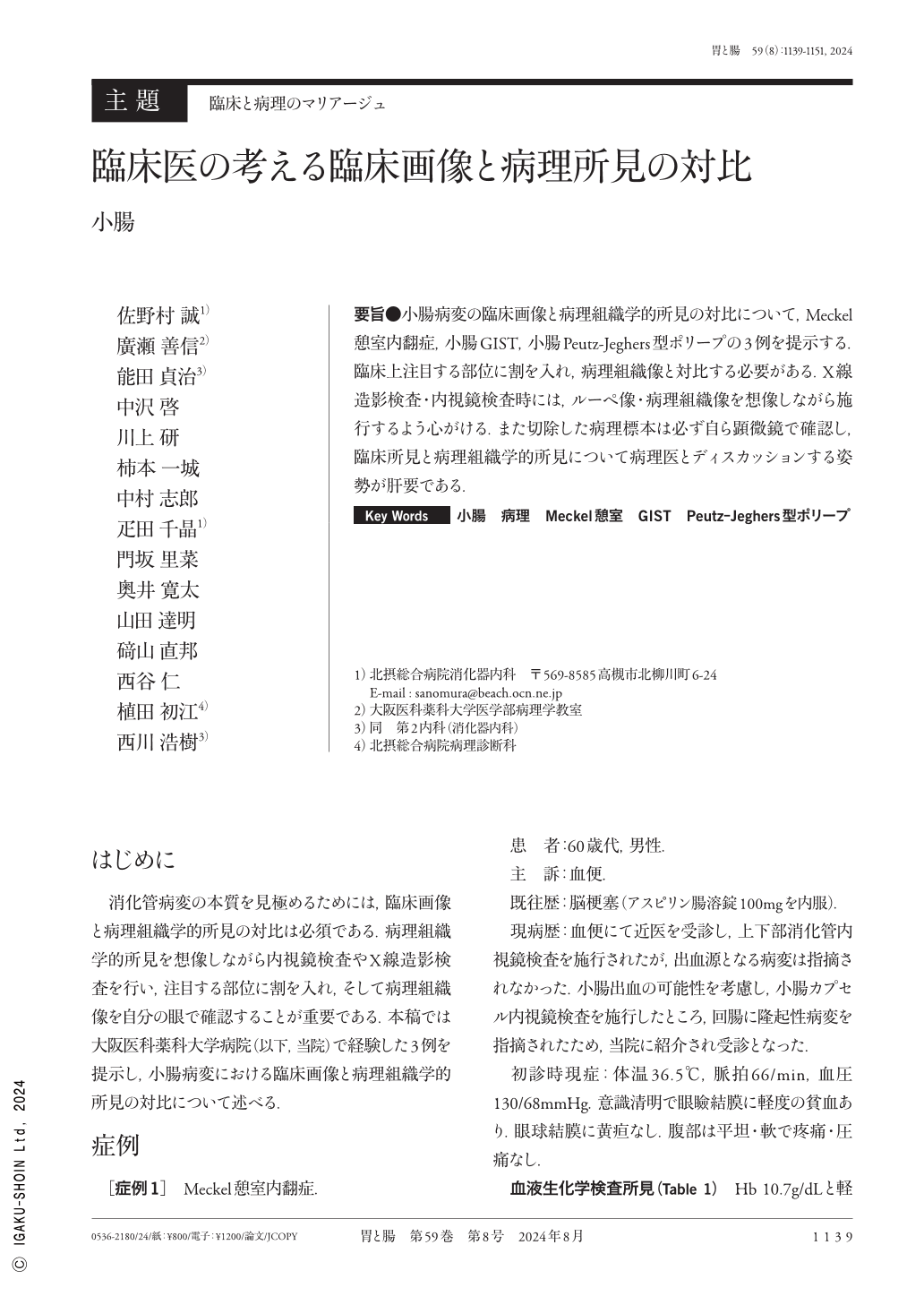
要旨●小腸病変の臨床画像と病理組織学的所見の対比について,Meckel憩室内翻症,小腸GIST,小腸Peutz-Jeghers型ポリープの3例を提示する.臨床上注目する部位に割を入れ,病理組織像と対比する必要がある.X線造影検査・内視鏡検査時には,ルーペ像・病理組織像を想像しながら施行するよう心がける.また切除した病理標本は必ず自ら顕微鏡で確認し,臨床所見と病理組織学的所見について病理医とディスカッションする姿勢が肝要である.
Three cases of small intestinal lesions—Meckel's diverticulum, small intestinal gastrointestinal stromal tumor, and Peutz−Jeghers-type polyps—are presented here to illustrate the contrast between clinical images and pathological findings. The clinical focus of each case should be created and compared with the histopathology. Additionally, cross-sectional and histopathological images should be analyzed during radiographic and endoscopic examinations. It is also crucial to examine the excised pathological specimen under a microscope and discuss the clinical and histopathological findings with a pathologist.
Copyright © 2024, Igaku-Shoin Ltd. All rights reserved.