Japanese
English
- 有料閲覧
PPV購入案内
1,320円 (1,200円+税10%) こちらは、884ページから892ページの文献です。購入には医書.jp本会員登録が必要です。
会員登録完了後に改めて上記「購入サイトへ」を選択してください。
または「購入サイトへ」選択後に「購入ログイン」、「新規会員登録」の順に進んでください。
会員登録について詳しくはをご確認ください。
- Abstract 文献概要
- 1ページ目 Look Inside
- 参考文献 Reference
- サイト内被引用 Cited by
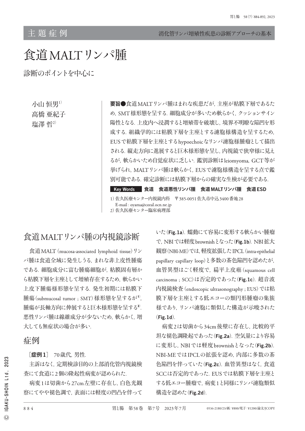
要旨●食道MALTリンパ腫はまれな疾患だが,主座が粘膜下層であるため,SMT様形態を呈する.細胞成分が多いため軟らかく,クッションサイン陽性となる.上皮内へ浸潤すると増殖帯を破壊し,境界不明瞭な陥凹を形成する.組織学的には粘膜下層を主座とする濾胞様構造を呈するため,EUSで粘膜下層を主座とするhypoechoicなリンパ濾胞様腫瘤として描出される.縦走方向に進展すると巨木様形態を呈し,内視鏡で狭窄様に見えるが,軟らかいため自覚症状に乏しい.鑑別診断はleiomyoma,GCT等が挙げられ,MALTリンパ腫は軟らかく,EUSで濾胞様構造を呈する点で鑑別可能である.確定診断には粘膜下層からの確実な生検が必要である.
Esophageal MALT(mucoid-associated lymphoid tissue)lymphoma is a rare disease. Since its main locus is the submucosa, it shows submucosal tumor-like morphology. It is a soft submucosal tumor with a positive cushion sign because of the several associated cellular components. When it infiltrates into the epithelium, it destroys the proliferative zone and forms an indistinct depression. Histologically, it is characterized by a lymph follicle-like structure. Therefore, the EUS(endoscopic ultrasound)indicates a lymph follicle-like hypoechoic mass in the submucosal layer. Additionally, when MALT lymphoma progresses in the longitudinal direction, it acquires a giant tree-like shape and appears like a stenosis on an endoscope ; nonetheless, it being a soft tumor, only a few subjective symptoms are presented. Differential diagnoses include leiomyoma and granular cell tumor ; however, MALT lymphoma can be differentiated based on its softness and lymph follicle-like structure observed on EUS. A biopsy of the submucosa is required for a definitive diagnosis.
Copyright © 2023, Igaku-Shoin Ltd. All rights reserved.