Japanese
English
- 有料閲覧
PPV購入案内
1,320円 (1,200円+税10%) こちらは、275ページから285ページの文献です。購入には医書.jp本会員登録が必要です。
会員登録完了後に改めて上記「購入サイトへ」を選択してください。
または「購入サイトへ」選択後に「購入ログイン」、「新規会員登録」の順に進んでください。
会員登録について詳しくはをご確認ください。
- Abstract 文献概要
- 1ページ目 Look Inside
- 参考文献 Reference
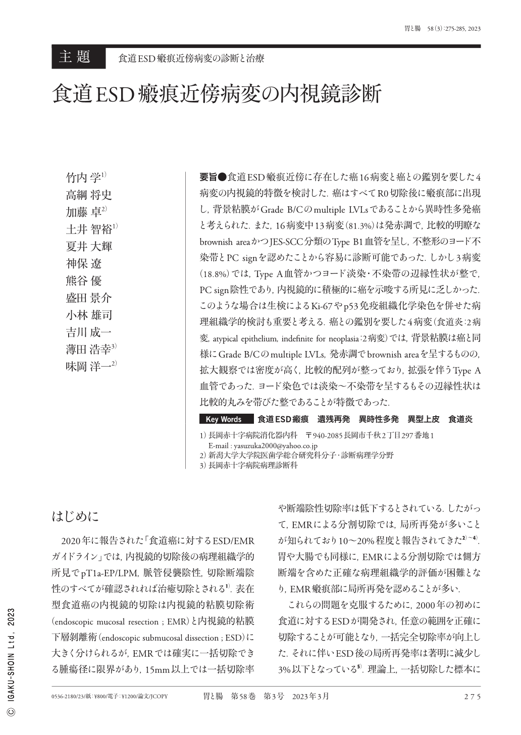
要旨●食道ESD瘢痕近傍に存在した癌16病変と癌との鑑別を要した4病変の内視鏡的特徴を検討した.癌はすべてR0切除後に瘢痕部に出現し,背景粘膜がGrade B/Cのmultiple LVLsであることから異時性多発癌と考えられた.また,16病変中13病変(81.3%)は発赤調で,比較的明瞭なbrownish areaかつJES-SCC分類のType B1血管を呈し,不整形のヨード不染帯とPC signを認めたことから容易に診断可能であった.しかし3病変(18.8%)では,Type A血管かつヨード淡染・不染帯の辺縁性状が整で,PC sign陰性であり,内視鏡的に積極的に癌を示唆する所見に乏しかった.このような場合は生検によるKi-67やp53免疫組織化学染色を併せた病理組織学的検討も重要と考える.癌との鑑別を要した4病変(食道炎:2病変,atypical epithelium,indefinite for neoplasia:2病変)では,背景粘膜は癌と同様にGrade B/Cのmultiple LVLs,発赤調でbrownish areaを呈するものの,拡大観察では密度が高く,比較的配列が整っており,拡張を伴うType A血管であった.ヨード染色では淡染〜不染帯を呈するもその辺縁性状は比較的丸みを帯びた整であることが特徴であった.
We evaluated the endoscopic features of the 16 ESCC(esophageal squamous cell carcinomas)and 4 lesions that needed differentiation with cancer, and they were in the vicinity of the esophagus endoscopic submucosal dissection scar. Due to R0 resection and the fact that the background mucosa had several LVLs(Lugol-voiding lesions)of Grade B/C and a high chance of metachronous multiple cancer, all malignant lesions were assumed to have metachronous cancer. In addition, 13(81.3%)among 16 lesions showed reddish color. Type B1 vessels of the JES-SCC classification in the relatively clear brownish area and positive PC(pink color)sign indicating iodine unstained lesion with an irregular shape like typical ESCC. However, 3 lesions(18.8%)demonstrated Type A vessels and negative PC sign with regular shaped unstained iodine or weak stained lesion. In these situations, it might be challenging to make a diagnosis of carcinoma simply on endoscopic observations, thus it is crucial to combine a biopsy with a pathological examination that includes p53 and Ki-67 immunostaining.
For 4 lesions(esophagitis:2, Atypical epithelium, indefinite for neoplasia:2)that required differentiation with cancer, we identified the following endoscopic characteristics:1)multiple LVLs of Grade B/C on background mucosa, 2)reddish in color and clear brownish area, 3)Type A vessels having high density with only dilatation and regular arrangement, and 4)regular shaped iodine unstained or weak stained lesion.
Copyright © 2023, Igaku-Shoin Ltd. All rights reserved.