Japanese
English
- 有料閲覧
- Abstract 文献概要
- 1ページ目 Look Inside
- 参考文献 Reference
- サイト内被引用 Cited by
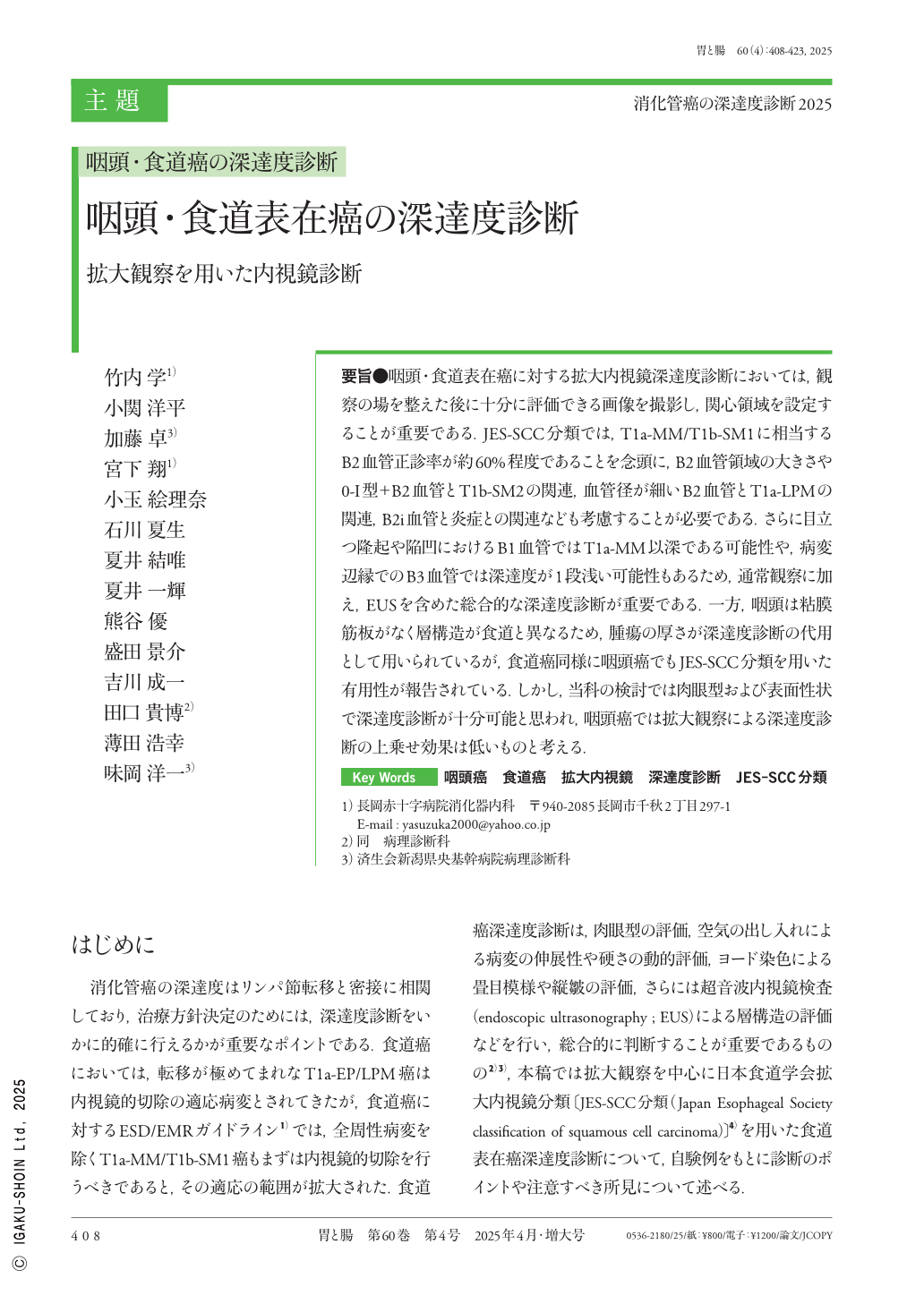
要旨●咽頭・食道表在癌に対する拡大内視鏡深達度診断においては,観察の場を整えた後に十分に評価できる画像を撮影し,関心領域を設定することが重要である.JES-SCC分類では,T1a-MM/T1b-SM1に相当するB2血管正診率が約60%程度であることを念頭に,B2血管領域の大きさや0-I型+B2血管とT1b-SM2の関連,血管径が細いB2血管とT1a-LPMの関連,B2i血管と炎症との関連なども考慮することが必要である.さらに目立つ隆起や陥凹におけるB1血管ではT1a-MM以深である可能性や,病変辺縁でのB3血管では深達度が1段浅い可能性もあるため,通常観察に加え,EUSを含めた総合的な深達度診断が重要である.一方,咽頭は粘膜筋板がなく層構造が食道と異なるため,腫瘍の厚さが深達度診断の代用として用いられているが,食道癌同様に咽頭癌でもJES-SCC分類を用いた有用性が報告されている.しかし,当科の検討では肉眼型および表面性状で深達度診断が十分可能と思われ,咽頭癌では拡大観察による深達度診断の上乗せ効果は低いものと考える.
In magnifying endoscopic assessment invasion depth in superficial carcinoma of the pharynx and esophagus, it is crucial to define the region of interest by capturing endoscopic images that allow for thorough evaluation after properly preparing the observation field. Based on the JES-SCC classification, the positive diagnosis rate of B2 vessels, which correspond to T1a-MM/T1b-SM1 is approximately 60%, Therefore, factors such as B2 vessel size, the relationship between 0-I+B2 vessels and T1b-SM2, the correlation between small-diameter B2 vessels and T1a-LPM, the association between B2i vessels and inflammation must be considered. Furthermore, B1 vessels in prominent protuberances and depressions may indicate a deeper invasion than T1a-MM, whereas B3 vessels at the lesion's edge may correspond to a shallower depth. However, because the pharynx has a different layered structure from the esophagus, the tumor thickness is used as a substitute for the invasion depth diagnosis. Our study, suggests that depth diagnosis based on gross tumor type and surface characteristics is generally sufficient, with magnification providing minimal additional value in assessing invasion depth in pharyngeal cancer.

Copyright © 2025, Igaku-Shoin Ltd. All rights reserved.


