Japanese
English
- 有料閲覧
- Abstract 文献概要
- 1ページ目 Look Inside
- 参考文献 Reference
- サイト内被引用 Cited by
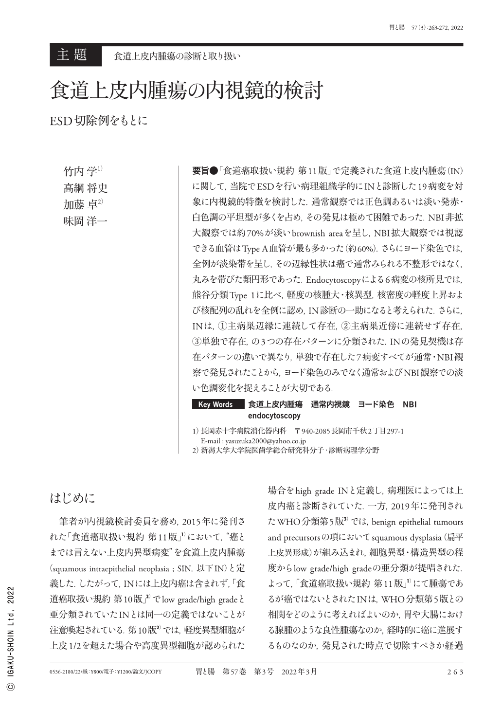
要旨●「食道癌取扱い規約 第11版」で定義された食道上皮内腫瘍(IN)に関して,当院でESDを行い病理組織学的にINと診断した19病変を対象に内視鏡的特徴を検討した.通常観察では正色調あるいは淡い発赤・白色調の平坦型が多くを占め,その発見は極めて困難であった.NBI非拡大観察では約70%が淡いbrownish areaを呈し,NBI拡大観察では視認できる血管はType A血管が最も多かった(約60%).さらにヨード染色では,全例が淡染帯を呈し,その辺縁性状は癌で通常みられる不整形ではなく,丸みを帯びた類円形であった.Endocytoscopyによる6病変の核所見では,熊谷分類Type 1に比べ,軽度の核腫大・核異型,核密度の軽度上昇および核配列の乱れを全例に認め,IN診断の一助になると考えられた.さらに,INは,①主病巣辺縁に連続して存在,②主病巣近傍に連続せず存在,③単独で存在,の3つの存在パターンに分類された.INの発見契機は存在パターンの違いで異なり,単独で存在した7病変すべてが通常・NBI観察で発見されたことから,ヨード染色のみでなく通常およびNBI観察での淡い色調変化を捉えることが大切である.
According to the 11th edition Japanese classification of Esophageal Cancer, esophageal squamous IN(intraepithelial neoplasia)is defined as a low-grade tumor excluding carcinoma in situ. Because endoscopic findings of IN are still unclear, we assessed the endoscopic features of 19 lesions, and two expert gastrointestinal pathologists identified the resected specimens as IN based on pathological findings, including immunostaining. Conventional endoscopic findings for IN primarily showed the same color as the background mucosa or light reddish and whitish color, and the diagnosis of IN by white-light imaging endoscopy was extremely difficult. Approximately 70% of IN showed a weak brownish area on non magnifying NBI(narrow band imaging), and Type A vessels according to the Japanese Esophageal Society's classification were the most common lesions in which vessels could be recognized visually(approximately 60%)on magnifying NBI. Iodine staining revealed that all lesions were mild and unstained, and the border characteristics were oval, not irregular as usually observed in cancer. In the nuclear findings from endocyotoscopy of six lesions, we detected slight nuclear swelling, dyskaryosis, increased nuclear density, and disordered nuclear sequence in all cases compared with Kumagai classification Type 1, and we considered that iodine aided the IN diagnosis. Further, we classified the IN existence pattern into the following categories:1)the main lesion in succession, 2)without continuing in the main lesion vicinity, and 3)independent. It differs in each existence pattern in a discovery opportunity, and IN must pay attention to light color changes by WLI and NBI observation, as well as iodine spraying all seven independent lesions without continuing in the vicinity of the main lesion.

Copyright © 2022, Igaku-Shoin Ltd. All rights reserved.


