Japanese
English
- 有料閲覧
PPV購入案内
1,320円 (1,200円+税10%) こちらは、501ページから514ページの文献です。購入には医書.jp本会員登録が必要です。
会員登録完了後に改めて上記「購入サイトへ」を選択してください。
または「購入サイトへ」選択後に「購入ログイン」、「新規会員登録」の順に進んでください。
会員登録について詳しくはをご確認ください。
- Abstract 文献概要
- 1ページ目 Look Inside
- 参考文献 Reference
- サイト内被引用 Cited by
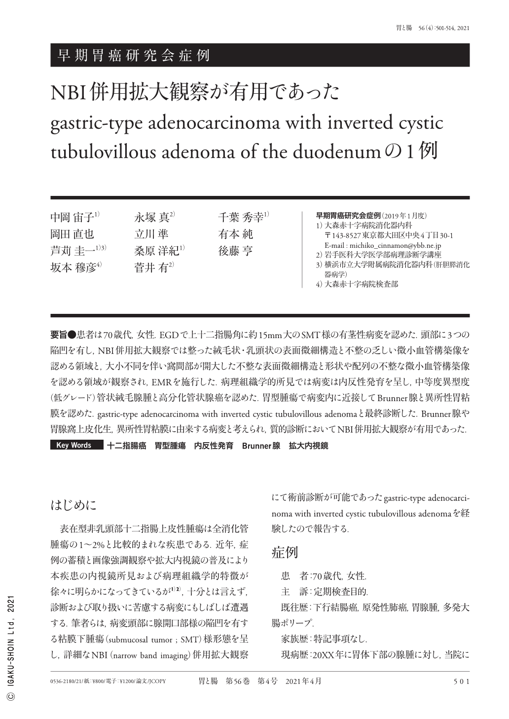
要旨●患者は70歳代,女性.EGDで上十二指腸角に約15mm大のSMT様の有茎性病変を認めた.頭部に3つの陥凹を有し,NBI併用拡大観察では整った絨毛状・乳頭状の表面微細構造と不整の乏しい微小血管構築像を認める領域と,大小不同を伴い窩間部が開大した不整な表面微細構造と形状や配列の不整な微小血管構築像を認める領域が観察され,EMRを施行した.病理組織学的所見では病変は内反性発育を呈し,中等度異型度(低グレード)管状絨毛腺腫と高分化管状腺癌を認めた.胃型腫瘍で病変内に近接してBrunner腺と異所性胃粘膜を認めた.gastric-type adenocarcinoma with inverted cystic tubulovillous adenomaと最終診断した.Brunner腺や胃腺窩上皮化生,異所性胃粘膜に由来する病変と考えられ,質的診断においてNBI併用拡大観察が有用であった.
A woman in her 70s was found to have an approximately 15-mm submucosal tumor-like pedunculated lesion on the superior duodenal angle, which was revealed by esophagogastroduodenoscopy. Three depressed sites were seen on the surface of the lesion, where magnifying narrow band imaging revealed characteristic areas with a regular villous and papillary microsurface pattern or regular microvessel construction and large intervening irregular microsurface pattern or microvessel construction. The lesion was finally diagnosed as carcinoma with adenoma and was subsequently treated by endoscopic mucosal resection. Histopathological findings revealed a moderate grade of atypia in the tubulovillous adenoma and a well-differentiated tubular adenocarcinoma. Moreover, the depressed areas had the characteristics of inverted growth. The lesion had a gastric phenotype, and the Brunner's gland and the ectopic gastric mucosa were found close to the lesion. Therefore, we confirmed the diagnosis as gastric-type adenocarcinoma with inverted cystic tubulovillous adenoma. The lesion was believed to have originated from the Brunner's gland, and intestinal metaplasia in the ectopic gastric mucosa, observed using magnifying narrow band imaging, was useful in the qualitative diagnosis.
Copyright © 2021, Igaku-Shoin Ltd. All rights reserved.