Japanese
English
- 有料閲覧
PPV購入案内
1,320円 (1,200円+税10%) こちらは、1485ページから1492ページの文献です。購入には医書.jp本会員登録が必要です。
会員登録完了後に改めて上記「購入サイトへ」を選択してください。
または「購入サイトへ」選択後に「購入ログイン」、「新規会員登録」の順に進んでください。
会員登録について詳しくはをご確認ください。
- Abstract 文献概要
- 1ページ目 Look Inside
- 参考文献 Reference
- サイト内被引用 Cited by
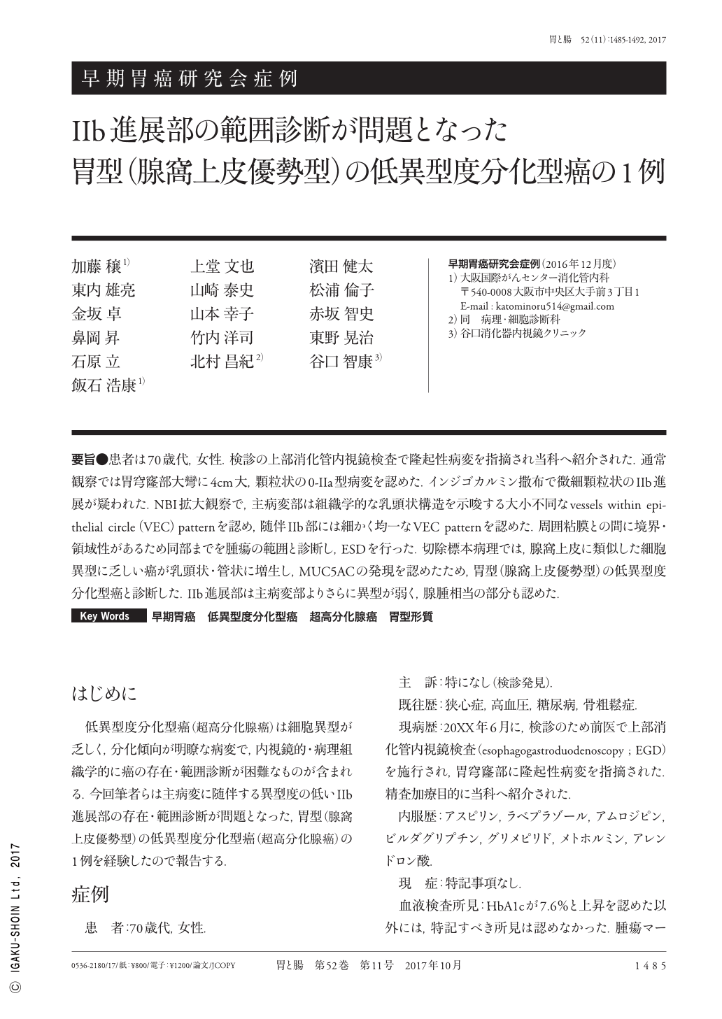
要旨●患者は70歳代,女性.検診の上部消化管内視鏡検査で隆起性病変を指摘され当科へ紹介された.通常観察では胃穹窿部大彎に4cm大,顆粒状の0-IIa型病変を認めた.インジゴカルミン撒布で微細顆粒状のIIb進展が疑われた.NBI拡大観察で,主病変部は組織学的な乳頭状構造を示唆する大小不同なvessels within epithelial circle(VEC)patternを認め,随伴IIb部には細かく均一なVEC patternを認めた.周囲粘膜との間に境界・領域性があるため同部までを腫瘍の範囲と診断し,ESDを行った.切除標本病理では,腺窩上皮に類似した細胞異型に乏しい癌が乳頭状・管状に増生し,MUC5ACの発現を認めたため,胃型(腺窩上皮優勢型)の低異型度分化型癌と診断した.IIb進展部は主病変部よりさらに異型が弱く,腺腫相当の部分も認めた.
A woman in her 70s was admitted to our hospital for further examination of abnormal gastric fundus findings detected by screening endoscopy. In WL(white light)images, a 40mm flat elevated lesion was observed in the great curvature of the gastric fundus. This lesion had a granular surface, and the lesion color was same as the surrounding normal mucosa. Chromoendoscopy with indigo carmine revealed a fine granular flat tumor extension, which was not clear in the WL image. Narrow-band imaging with magnification visualized VEC(vessels within an epithelial circle)pattern in the lesion, which was characteristic of a histological papillary structure. The VEC pattern observed in the elevated part of the lesion was irregular in size, whereas the VEC pattern observed in the flat extending part of the lesion was small and uniform. Endoscopic submucosal dissection was performed, and histological findings of the resected tumor consisted of columnar cells with mild nuclear atypia, which resembled foveolar cells, showing papillary/tubular projections. Immunohistochemically, the tumor glands were strongly positive for MUC5AC. A gastric-type low-grade differentiated-type adenocarcinoma(foveolar predominant-type neoplasia)was diagnosed. The degree of nuclear atypia of the accompanied flat component, which was unclear in the WL image, was lower than that of the elevated part of the lesion.
Copyright © 2017, Igaku-Shoin Ltd. All rights reserved.