Japanese
English
- 有料閲覧
- Abstract 文献概要
- 1ページ目 Look Inside
- 参考文献 Reference
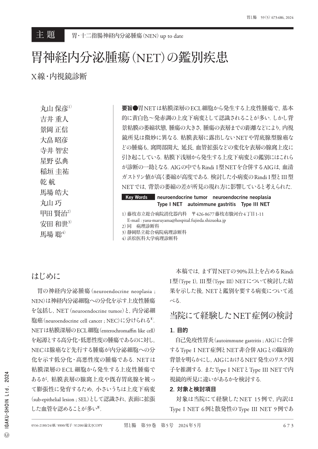
要旨●胃NETは粘膜深層のECL細胞から発生する上皮性腫瘍で,基本的に黄白色〜発赤調の上皮下病変として認識されることが多い.しかし背景粘膜の萎縮状態,腫瘍の大きさ,腫瘍の表層までの距離などにより,内視鏡所見は微妙に異なる.粘膜表層に露出しないNETや胃底腺型腺癌などの腫瘍も,窩間部開大,延長,血管拡張などの変化を表層の腺窩上皮に引き起こしている.粘膜下浅層から発生する上皮下病変との鑑別にはこれらが診断の一助となる.AIGの中でもRindi I型NETを合併するAIGは,血清ガストリン値が高く萎縮が高度である.検討した小病変のRindi I型とIII型NETでは,背景の萎縮の差が所見の現れ方に影響していると考えられた.
Gastric NETs(neuroendocrine tumors)are epithelial tumors that arise from enterochromaffin-like cells in the deep mucosal layer and are generally recognized as yellowish-white to reddish subepithelial lesions. However, endoscopic findings vary slightly depending on factors such as the state of atrophy of the background mucosa, the tumor size, and the distance to the tumor surface. Tumors that are not exposed to the mucosal surface, such as NETs and fundic gland-type adenocarcinoma, also induce superficial crypt epithelium changes, such as foveal enlargement, elongation, and vascular dilation. This aids in diagnosis especially when differentiating it from subepithelial lesions that arise from the shallow submucosal layer. Among AIGs, AIG associated with Rindi type I NET presents with high serum gastrin levels and severe atrophy. The difference in background atrophy between the small lesions of Rindi type I and type III NETs was believed to influence the morphology of the findings.

Copyright © 2024, Igaku-Shoin Ltd. All rights reserved.


