Japanese
English
- 有料閲覧
PPV購入案内
1,320円 (1,200円+税10%) こちらは、217ページから224ページの文献です。購入には医書.jp本会員登録が必要です。
会員登録完了後に改めて上記「購入サイトへ」を選択してください。
または「購入サイトへ」選択後に「購入ログイン」、「新規会員登録」の順に進んでください。
会員登録について詳しくはをご確認ください。
- Abstract 文献概要
- 1ページ目 Look Inside
- 参考文献 Reference
- サイト内被引用 Cited by
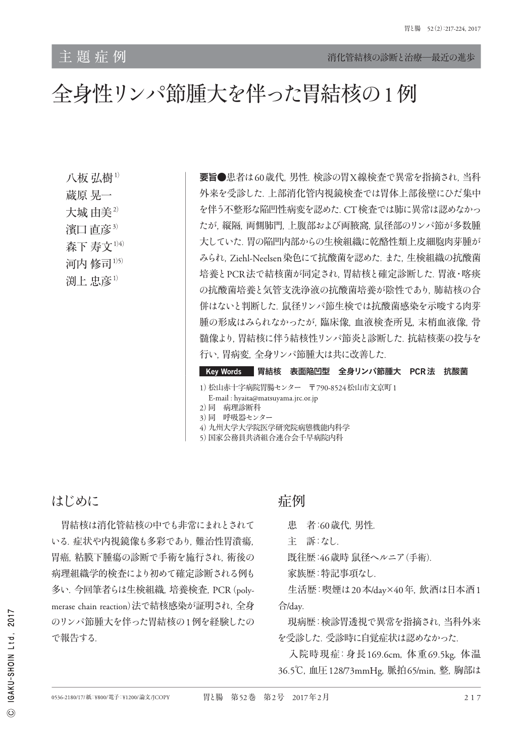
要旨●患者は60歳代,男性.検診の胃X線検査で異常を指摘され,当科外来を受診した.上部消化管内視鏡検査では胃体上部後壁にひだ集中を伴う不整形な陥凹性病変を認めた.CT検査では肺に異常は認めなかったが,縦隔,両側肺門,上腹部および両腋窩,鼠径部のリンパ節が多数腫大していた.胃の陥凹内部からの生検組織に乾酪性類上皮細胞肉芽腫がみられ,Ziehl-Neelsen染色にて抗酸菌を認めた.また,生検組織の抗酸菌培養とPCR法で結核菌が同定され,胃結核と確定診断した.胃液・喀痰の抗酸菌培養と気管支洗浄液の抗酸菌培養が陰性であり,肺結核の合併はないと判断した.鼠径リンパ節生検では抗酸菌感染を示唆する肉芽腫の形成はみられなかったが,臨床像,血液検査所見,末梢血液像,骨髄像より,胃結核に伴う結核性リンパ節炎と診断した.抗結核薬の投与を行い,胃病変,全身リンパ節腫大は共に改善した.
A 60-year-old asymptomatic man was referred to our hospital for the evaluation of a gastric lesion detected using fluorography of the stomach in a medical checkup. EGD(esophagogastroduodenoscopy)showed an irregularly shaped depressed lesion with converging folds on the posterior wall of the upper gastric corpus. Fluorine- 18-fluorodeoxyglucose positron emission tomography showed markedly increased accumulation in the lymph nodes of the mediastinum, pulmonary hilum, and upper abdomen. Chest computed tomography revealed no evidence of pulmonary tuberculosis. Biopsy of the lesion showed granulomatous inflammation with caseation necrosis and Langerhans giant cells. Acid-fast bacilli were detected using both Ziehl-Neelsen staining and mycobacterium culture. Polymerase chain reaction test for tuberculosis was also positive. Biopsy of the inguinal lymph node revealed nonspecific inflammation without any neoplastic cells or granulomas. The patient was diagnosed as having gastric tuberculosis with systemic lymphadenopathy and subsequently underwent antituberculous treatment. Six months later, the gastric lesion and lymphadenopathy were resolved.
Copyright © 2017, Igaku-Shoin Ltd. All rights reserved.