Japanese
English
- 有料閲覧
PPV購入案内
1,320円 (1,200円+税10%) こちらは、1185ページから1196ページの文献です。購入には医書.jp本会員登録が必要です。
会員登録完了後に改めて上記「購入サイトへ」を選択してください。
または「購入サイトへ」選択後に「購入ログイン」、「新規会員登録」の順に進んでください。
会員登録について詳しくはをご確認ください。
- Abstract 文献概要
- 1ページ目 Look Inside
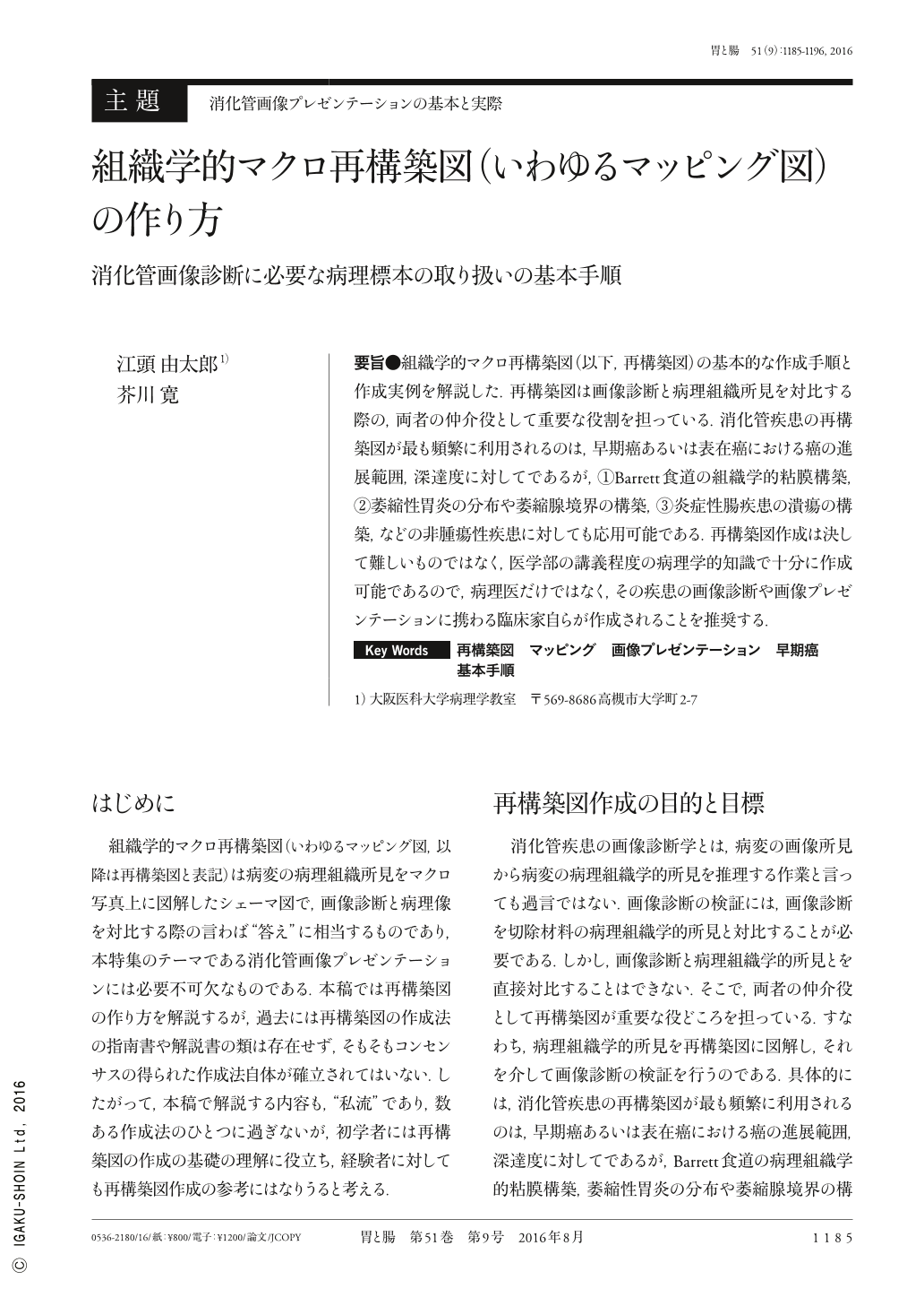
要旨●組織学的マクロ再構築図(以下,再構築図)の基本的な作成手順と作成実例を解説した.再構築図は画像診断と病理組織所見を対比する際の,両者の仲介役として重要な役割を担っている.消化管疾患の再構築図が最も頻繁に利用されるのは,早期癌あるいは表在癌における癌の進展範囲,深達度に対してであるが,①Barrett食道の組織学的粘膜構築,②萎縮性胃炎の分布や萎縮腺境界の構築,③炎症性腸疾患の潰瘍の構築,などの非腫瘍性疾患に対しても応用可能である.再構築図作成は決して難しいものではなく,医学部の講義程度の病理学的知識で十分に作成可能であるので,病理医だけではなく,その疾患の画像診断や画像プレゼンテーションに携わる臨床家自らが作成されることを推奨する.
The basic procedure for reconstructing histological macro images(reconstructed images)has been explained with actual examples. Reconstructed images play a key role as an intermediary when comparing diagnostic imaging results with histopathological findings. Reconstructed images of gastrointestinal disease are most frequently used to determine the extent and depth of disease progression in early-stage and superficial cancer. However, they can also be used for non-neoplastic diseases such as 1)the histological mucosal structure in Barrett's esophagus, 2)distribution of atrophic gastritis and structure of the atrophic gland border, and 3)structure of ulceration in inflammatory bowel disease. Reconstructed images are not difficult to create. Because images can be adequately created with medical school lecture-level knowledge of pathology, we recommend that they be created not only by pathologists but also by clinicians who have performed the diagnostic imaging for such diseases or are involved in image presentations.
Copyright © 2016, Igaku-Shoin Ltd. All rights reserved.