Japanese
English
- 有料閲覧
PPV購入案内
1,320円 (1,200円+税10%) こちらは、588ページから593ページの文献です。購入には医書.jp本会員登録が必要です。
会員登録完了後に改めて上記「購入サイトへ」を選択してください。
または「購入サイトへ」選択後に「購入ログイン」、「新規会員登録」の順に進んでください。
会員登録について詳しくはをご確認ください。
- Abstract 文献概要
- 1ページ目 Look Inside
- 参考文献 Reference
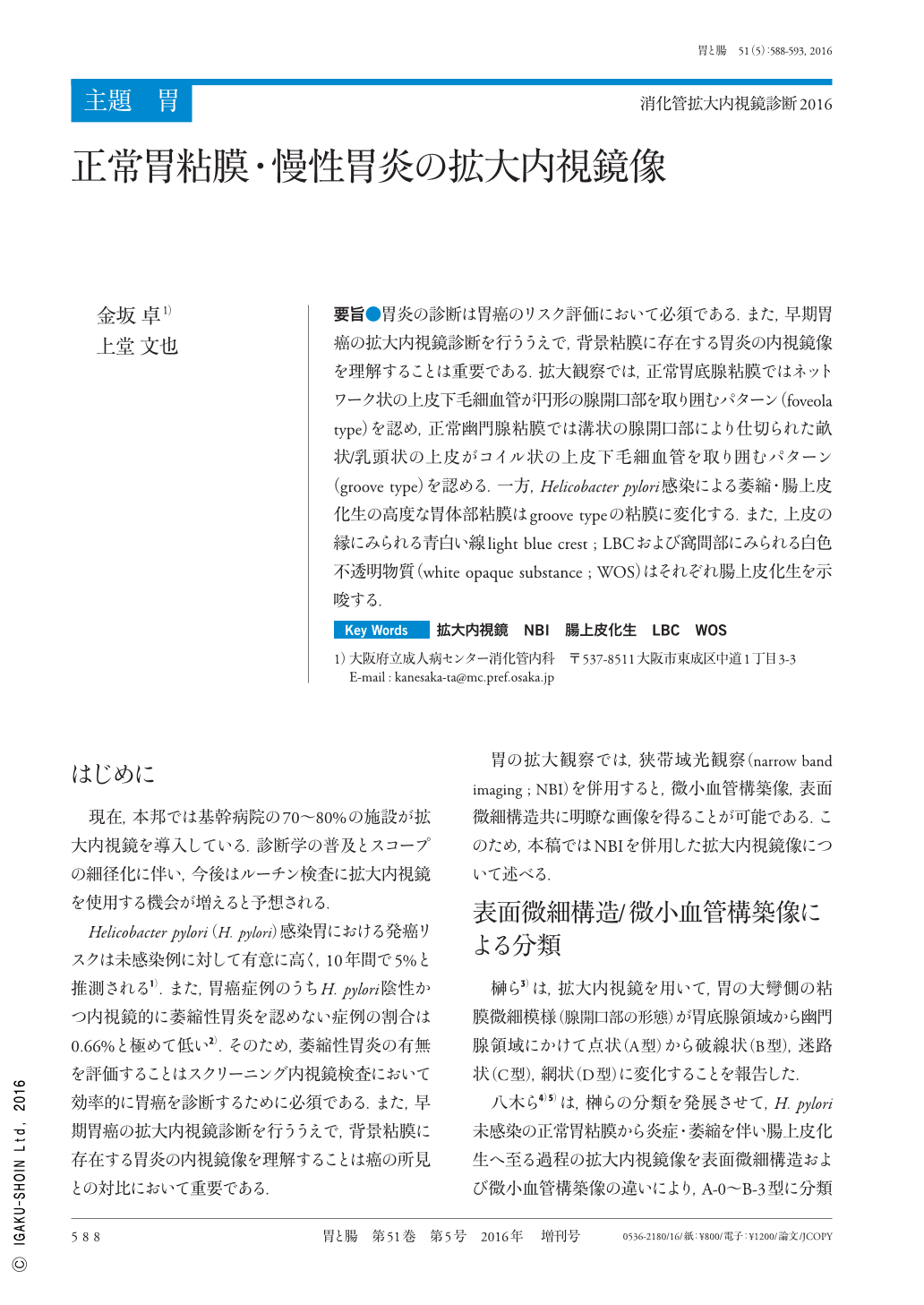
要旨●胃炎の診断は胃癌のリスク評価において必須である.また,早期胃癌の拡大内視鏡診断を行ううえで,背景粘膜に存在する胃炎の内視鏡像を理解することは重要である.拡大観察では,正常胃底腺粘膜ではネットワーク状の上皮下毛細血管が円形の腺開口部を取り囲むパターン(foveola type)を認め,正常幽門腺粘膜では溝状の腺開口部により仕切られた畝状/乳頭状の上皮がコイル状の上皮下毛細血管を取り囲むパターン(groove type)を認める.一方,Helicobacter pylori感染による萎縮・腸上皮化生の高度な胃体部粘膜はgroove typeの粘膜に変化する.また,上皮の縁にみられる青白い線light blue crest ; LBCおよび窩間部にみられる白色不透明物質(white opaque substance ; WOS)はそれぞれ腸上皮化生を示唆する.
Evaluating the prevalence of gastritis by magnifying endoscopy is useful for estimating the risk of gastric cancer. Moreover, understanding the endoscopic appearances of gastritis is important for diagnosing gastric cancer using magnifying endoscopy.
Regular arrangement of collecting venules and round crypt openings that are surrounded by a network of sub-epithelial capillaries represent predictive appearances of a normal corpus mucosa that is negative for Helicobacter pylori. In contrast, normal antral mucosa shows a ridged/papillary micro-surface pattern that encases coiled sub-epithelial capillaries. As atrophic gastritis progresses, the corpus mucosa, exhibiting round crypt openings, transforms into a ridged/papillary mucosa that is similar to the antral mucosa. Identification of a ridged/papillary mucosa with a light blue crest or white opaque substance represents predictive appearances of intestinal metaplasia.
Copyright © 2016, Igaku-Shoin Ltd. All rights reserved.