Japanese
English
- 有料閲覧
PPV購入案内
1,320円 (1,200円+税10%) こちらは、1147ページから1155ページの文献です。購入には医書.jp本会員登録が必要です。
会員登録完了後に改めて上記「購入サイトへ」を選択してください。
または「購入サイトへ」選択後に「購入ログイン」、「新規会員登録」の順に進んでください。
会員登録について詳しくはをご確認ください。
- Abstract 文献概要
- 1ページ目 Look Inside
- 参考文献 Reference
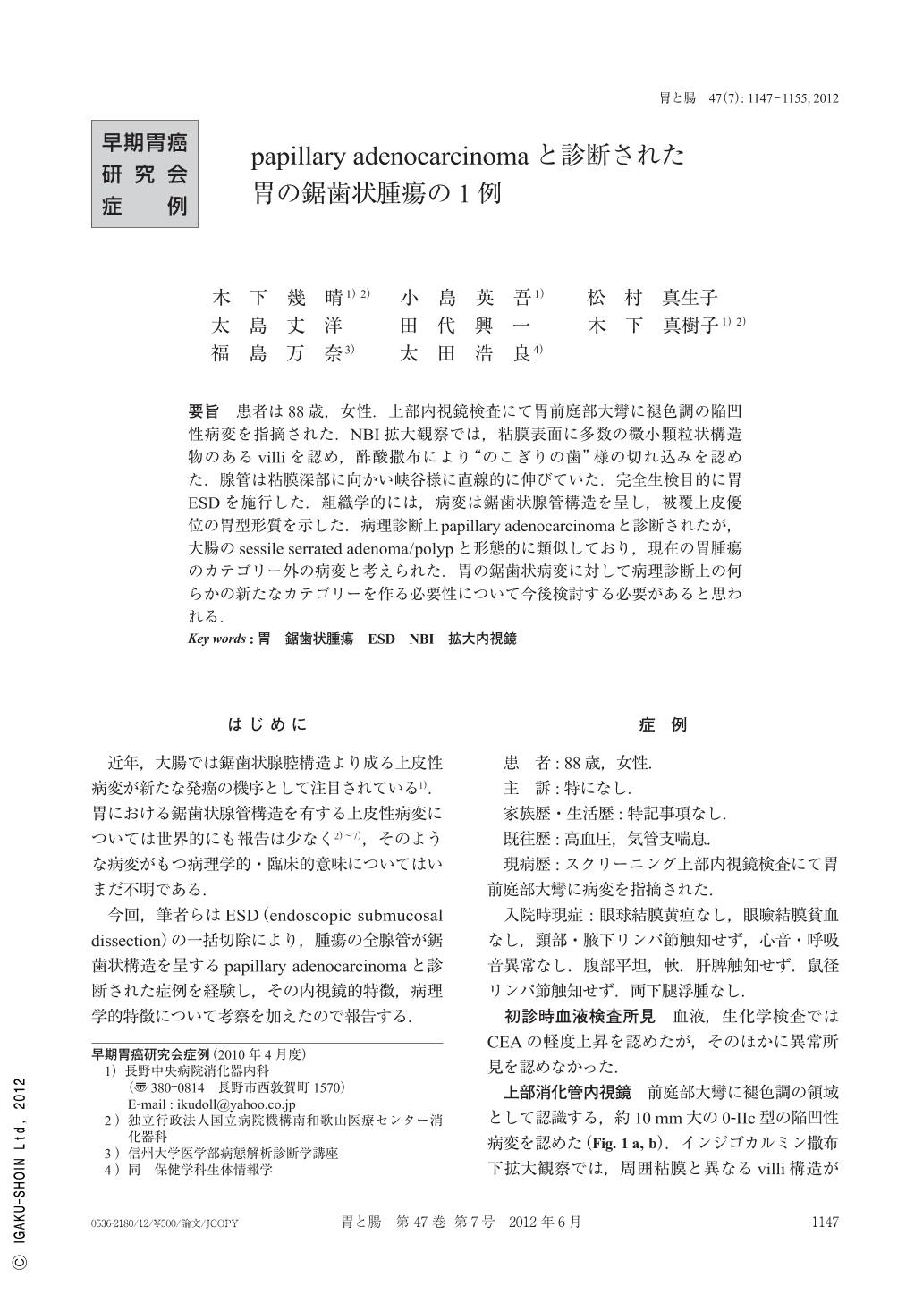
要旨 患者は88歳,女性.上部内視鏡検査にて胃前庭部大彎に褪色調の陥凹性病変を指摘された.NBI拡大観察では,粘膜表面に多数の微小顆粒状構造物のあるvilliを認め,酢酸撒布により“のこぎりの歯”様の切れ込みを認めた.腺管は粘膜深部に向かい峡谷様に直線的に伸びていた.完全生検目的に胃ESDを施行した.組織学的には,病変は鋸歯状腺管構造を呈し,被覆上皮優位の胃型形質を示した.病理診断上papillary adenocarcinomaと診断されたが,大腸のsessile serrated adenoma/polypと形態的に類似しており,現在の胃腫瘍のカテゴリー外の病変と考えられた.胃の鋸歯状病変に対して病理診断上の何らかの新たなカテゴリーを作る必要性について今後検討する必要があると思われる.
An 88-year-old woman was found, by upper endoscopy, to have a discolored depressed lesion in the greater curvature of the gastric antrum. NBI magnifying observation revealed many fine granular structures on the mucosal surface of the lesion, and acetic acid spraying showed slits shaped like“a saw tooth”. The glandular ducts showed a valley-like linear extension to the mucosal depth. Gastric ESD was performed for a complete biopsy. Histologically, the lesion showed a serrated gland structure, which was a predominantly gastric surface epithelial type. Although the lesion was pathologically diagnosed as papillary adenocarcinoma, it was morphologically similar to a sessile serrated adenoma/polyp in the large intestine, and was considered outside the current categories of gastric tumors. The need for creating some new pathological categories for gastric serrated lesions requires consideration in the future.
Copyright © 2012, Igaku-Shoin Ltd. All rights reserved.